

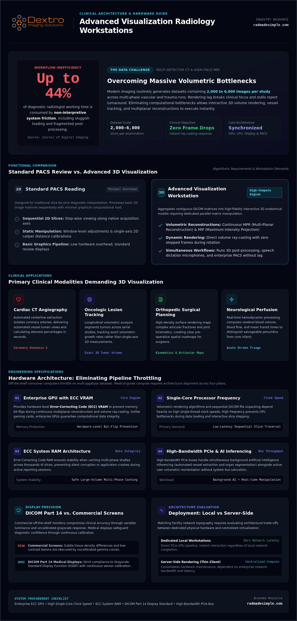
Studies published in the Journal of Digital Imaging reveal that diagnostic radiologists spend up to 44% of their working time on non-interpretive system friction, frequently waiting on sluggish dataset loading and fragmented post-processing applications. When you routinely interpret multi-phase vascular or trauma studies containing thousands of slices, rendering lag breaks clinical focus and stalls report turnaround. Selecting the right advanced visualization workstation for radiology means eliminating these computational bottlenecks so 3D volume rendering, vessel tracking, and multiplanar reconstructions execute instantly.
You already know that diagnostic velocity requires more than raw compute power; high-performance rendering hardware, calibrated medical displays, and PACS software must synchronize seamlessly. This 2026 buyer guide shows you how to architect, specify, and deploy an advanced visualization radiology workstation that accelerates post-processing workflows and sharpens diagnostic precision without inflating infrastructure costs. We examine optimal processing architectures, DICOM Part 14 display standards, and integration strategies to help you build a reliable, future-proof reading environment.
Key Takeaways
- Learn how a purpose-built advanced visualization workstation for radiology eliminates processing bottlenecks across massive volumetric CT, MRI, and PET datasets.
- Identify the optimal balance of enterprise GPU memory, single-core processor speed, and ECC system memory needed to accelerate complex 3D post-processing.
- Understand why commercial screens compromise clinical accuracy and how medical displays safeguard diagnostic confidence through continuous DICOM Part 14 calibration.
- Evaluate the architectural trade-offs between local workstation power and server-side rendering to match your facility's network topography.
- Apply a structured procurement framework that ensures seamless integration between reading hardware, specialized diagnostic monitors, and enterprise PACS software.
What Defines an Advanced Visualization Workstation for Modern Radiology?
An advanced visualization workstation for radiology is a dedicated compute engine designed for complex volumetric post-processing. While standard review displays present individual 2D image slices, an advanced workstation aggregates contiguous DICOM matrices into high-fidelity, interactive 3D anatomical models. Multi-detector CT and high-field MRI scans frequently produce datasets exceeding 2,000 to 6,000 images per study. Navigating these immense volumetric runs requires low-latency manipulation. Hardware stability directly governs your diagnostic velocity; eliminating frame drops and rendering pauses keeps radiologist reporting moving without interruption.
Historically, diagnostic environments routed raw data through standard picture archiving and communication systems (PACS) for static slice reviews. Today's clinical throughput requires deeper computational interaction. Advanced post-processing unlocks diagnostic evidence that static views obscure, ensuring radiologists quickly detect vascular stenosis, occult fractures, and organ lesions.
Primary Clinical Modalities Demanding 3D Visualization
Modern advanced visualization tools streamline multi-specialty workflows across several high-volume imaging domains:
- Cardiac CT Angiography: Automated centerline extraction isolates coronary arteries, providing automated vessel lumen views and calculating stenosis percentages in seconds.
- Oncologic Lesion Tracking: Longitudinal volumetric analysis segments tumors across serial studies, tracking exact volumetric growth rates rather than relying solely on single-axis 2D measurements.
- Orthopedic Surgical Planning: High-density surface rendering maps complex articular fractures and joint kinematics, giving orthopedic surgeons clear pre-operative spatial roadmaps.
- Neurological Perfusion: Real-time hemodynamic processing computes cerebral blood volume, blood flow, and mean transit times to distinguish salvageable penumbra from infarcted core tissue during acute stroke triage.
Advanced Post-Processing vs. Standard PACS Reading
Standard PACS software focuses on sequential 2D slice presentation, window-level adjustments, and basic distance calibrations. These basic operations demand minimal processing overhead. An advanced visualization workstation for radiology, by contrast, executes continuous multi-planar reconstruction (MPR), maximum intensity projection (MIP), and direct volume rendering.
These algorithmic techniques require dedicated GPU pipelines capable of parallel floating-point matrix manipulation. Radiologists rotating a 3D vascular volume need instant ray-casting updates, not stepped frames. Integrating these compute capabilities into purpose-built radiology reading workstations ensures that post-processing applications, dictation microphones, and enterprise PACS run simultaneously without lag.
Hardware Architecture: Essential Compute and GPU Specifications
Medical imaging algorithms also increasingly execute background artificial intelligence inferencing alongside active user interactions. Your physical architecture must handle simultaneous automated vessel extraction, organ segmentation, and real-time volumetric reorientation without exhausting system buses.
GPU Acceleration and Memory Bandwidth for Volumetric Reconstruction
Enterprise graphic processing units serve as the core rendering engine for intensive 3D post-processing. Unlike commercial gaming cards, enterprise medical GPUs provide hardware-level Error-Correcting Code (ECC) VRAM, preventing memory bit-flips that could introduce visual artifacts into diagnostic reconstructions.
- VRAM Capacity: Equipping workstations with 20 GB to 48 GB of GDDR6 ECC memory ensures that multi-phase contrast studies reside entirely on-card, eliminating rendering stutter.
- Compute Pipelines: Thousands of dedicated CUDA and OpenCL cores accelerate ray-casting algorithms and cinematic volume rendering at native screen resolutions.
- Interface Precision: Specialized medical display controllers deliver 10-bit and 12-bit grayscale outputs, which support compliance with the AAPM Task Group Report 270 display quality assurance guidelines by maintaining precise luminance response curves.
CPU Selection, System RAM, and High-Throughput NVMe Pipelines
Processor clock speed directly dictates user interface fluidity. While volumetric background rendering scales across multiple GPU cores, interactive MPR reformatting and slice indexing rely heavily on high single-threaded CPU frequencies. Selecting processors like modern Intel Xeon W-series or AMD Ryzen Threadripper PRO lines provides the high clock rates needed for responsive navigation alongside expansive PCIe lane configurations.
System memory must be sized appropriately to hold uncompressed DICOM volumes. Enterprise systems require a baseline of 64 GB to 128 GB of DDR5 ECC memory to eliminate memory paging bottlenecks when multiple comparative prior studies remain open. Storage architecture demands PCIe 4.0 or PCIe 5.0 NVMe solid-state drives operating at sequential read speeds exceeding 7,000 MB/s. These high-throughput pipelines pull multi-gigabyte studies into active memory within fractions of a second, ensuring uninterrupted review during demanding teleradiology shifts.
If you need purpose-built systems pre-configured to meet these computational demands, explore turnkey diagnostic hardware solutions tailored for hospital networks and imaging centers.
Display Integration: Medical Diagnostic Monitors and DICOM Part 14
High-performance computing hardware delivers real clinical value only when paired with an equally capable medical display stack. An advanced visualization workstation for radiology generates intricate, high-contrast volumetric reconstructions that commercial office displays simply cannot reproduce accurately. Consumer-grade monitors prioritize dynamic color saturations and aggressive energy-saving dimming over optical linearity. When examining subtle parenchymal ground glass opacities or low-contrast liver lesions, deploying consumer screens risks missing early pathological indicators and invites severe regulatory non-compliance.
Treating monitors and processing units as separate purchasing silos undermines overall system performance. Diagnostic visualization requires hardware-level synchronization between the graphics card lookup tables and the monitor's internal optical sensors to ensure continuous, reproducible presentation across every reading session.
DICOM Grayscale Standard Display Function (GSDF) Compliance
The DICOM Part 14 GSDF standard establishes a mathematically linear relationship between pixel digital driving levels and perceived brightness across human vision. Maintaining this curve demands consistent minimum luminance (L'min ≥ 1.0 cd/m²) and maximum calibrated luminance (L'max ≥ 350 cd/m²), holding luminance response strictly within a ±10% tolerance band.
Because LED backlights degrade over time, medical monitors utilize integrated front-facing optical sensors that execute automated constancy checks in the background. Deploying certified solutions through Dextro Imaging Solutions provides automated, hands-free GSDF compliance across your fleet, correcting for backlight decay and ambient room luminance without requiring manual photometer calibration.
Resolution Thresholds and Multi-Modality Color Accuracy
Primary diagnostic interpretation requires certified resolution thresholds matched directly to modality demands:
- Cross-Sectional Imaging: Standard CT, MRI, and multi-slice post-processing require a minimum of 3MP to 5MP native matrix panels to prevent downsampling artifacts.
- Fused Multi-Modality Studies: PET/CT oncology overlays and multiparametric MRI maps rely on 10-bit or 12-bit native color rendering driven by internal 14-bit to 16-bit 3D lookup tables, eliminating digital banding along critical tissue margins.
- Multi-Head Calibration: Dual-head viewing environments demand strict color temperature and luminance matching between adjacent panels to eliminate visual fatigue during long interpretive shifts.
Establishing unified quality control across your visual and compute assets protects patient outcomes while fulfilling strict accreditation mandates. Review your ongoing facility audit procedures using this practical 2026 radiology equipment compliance checklist.
Deployment Architecture: Dedicated Workstations vs. Server-Side Rendering
Clinical leaders choosing an advanced visualization workstation for radiology must decide between dedicated local processing units and virtualized server-side rendering (SSR). While centralized server rendering promises simplified IT maintenance, it introduces significant network dependencies and recurring software overhead. Dedicated physical workstations eliminate network latency risks by keeping heavy graphic processing directly at the reading desk, ensuring uncompromising performance for high-throughput diagnostic shifts.
Hybrid architectures often provide the most practical middle ground. Diagnostic teams pair local workstations featuring dedicated compute hardware with secure, centralized archives, pulling full-fidelity DICOM datasets locally for uninterrupted manipulation. For facilities scaling centralized computational clusters or deep-learning imaging models, partnering with enterprise hardware specialists like E-Circles LLC provides the requisite AI infrastructure and high-performance GPU server solutions.
The Clinical Case for Dedicated Local Workstations
Dedicated local workstations provide predictable, hardware-direct rendering power. Because raw graphics pipelines execute locally on enterprise GPUs, radiologists experience zero frame-rate stutter during intensive 3D rotations, vascular centerline sculpting, or virtual fly-through procedures.
- Artifact-Free Image Quality: Streaming protocols rely on lossy compression codecs like H.264 or AV1 to transmit display pixels across local networks. Dedicated local engines display uncompressed volumetric matrices, eliminating compression artifacts that could obscure subtle microcalcifications or low-contrast liver lesions.
- Operational Independence: Local systems maintain full diagnostic functionality even during network slowdowns, archive server maintenance, or bandwidth throttling.
- Predictable Hardware Costs: Upgrading local client workstations avoids the complex software stack licensing and recurring hypervisor subscription fees required by virtualized GPU server pools.
Server-Side Thin Clients and Network Infrastructure Realities
Server-side thin clients process 3D volumes on centralized data-center clusters and stream interactive video feeds back to the user interface. This architecture simplifies software distribution and enables reading across standard enterprise laptops, but it demands strict network infrastructure to succeed. A sudden dip in network throughput introduces interactive lag, turning smooth 3D vessel manipulation into a stepped, delayed experience.
Server virtualization also carries heavy financial overhead. Enterprise server-side GPU slicing licenses, annual virtual desktop infrastructure (VDI) broker subscriptions, and required 10 GbE network overhauls can rapidly escalate total expenditure for independent practices. To accurately assess deployment expenses across both physical and virtual models, review our detailed radiology PACS pricing guide.
To evaluate which deployment architecture best aligns with your facility's study volume and IT network topography, consult the deployment specialists at Dextro Imaging Solutions for a custom hardware evaluation.
Evaluating and Specifying Your Workstation: A 5-Step Procurement Framework
Procuring an advanced visualization workstation for radiology requires a structured evaluation rather than assembling piecemeal parts. Disconnects between graphical processing power, diagnostic displays, and dictation peripherals create frustrating workflow bottlenecks. Following an objective procurement framework ensures your hardware deployment accelerates clinical turnaround, protects regulatory compliance, and simplifies multi-year life-cycle management.
Standardizing reading station footprints across your organization also minimizes ongoing IT maintenance friction, letting clinical teams move between desks without facing mismatched user interfaces or configuration errors.
Step-by-Step Diagnostic Workstation Specification Guide
Deploying a dependable reading environment requires working through five operational checkpoints:
- Audit Clinical Caseloads: Profile your modality mix and examine peak slice counts across cardiology, oncology, and neurology to establish realistic memory and processing baselines.
- Size Compute and Graphics Power: Select enterprise GPUs with sufficient ECC VRAM to hold entire volumetric series in active graphics memory alongside high base-clock processors.
- Specify Calibrated Displays: Pair graphics controllers with certified medical monitors that guarantee automated DICOM Part 14 adherence and meet required 3MP to 5MP diagnostic resolution thresholds.
- Validate Workflow Interoperability: Confirm certified driver compatibility across your enterprise PACS platform, specialized 3D post-processing suites, and clinical dictation software.
- Schedule Compliance and Maintenance: Establish proactive hardware support agreements and automate display constancy audits to satisfy ongoing quality standards without manual intervention.
Ergonomics, Acoustic Control, and Turnkey System Integration
Continuous volumetric processing generates substantial thermal energy. In quiet reading rooms, high-RPM cooling fans produce intrusive acoustic whine that elevates stress and impairs concentration during grueling reading shifts. Purpose-built systems mitigate this problem through acoustic chassis dampening, liquid cooling loops, or silent, high-static-pressure fans designed to keep noise levels below ambient room thresholds.
Turnkey Dextro Reading Stations deliver pre-configured, acoustically optimized hardware engineered specifically for intensive rendering and PACS stability. Complementing these compute engines with integrated RadVoice dictation tools unites high-resolution 3D reconstruction, image archiving, and immediate voice reporting into a unified, friction-free clinical workspace.
Optimize Your Reading Environment for Next-Generation Visualization
Modern volumetric imaging demands hardware that keeps pace with clinical complexity. Deploying the right advanced visualization workstation for radiology eliminates post-processing latency, safeguards continuous DICOM Part 14 calibration, and keeps radiologists focused on diagnostic precision rather than interface delays. When your compute architecture, medical displays, and PACS synchronize seamlessly, your entire imaging service operates with greater confidence and speed.
Achieving this level of reliability requires moving past piecemeal component sourcing. Equip your department with high-performance Dextro Reading Stations to provide your clinical teams with turnkey systems optimized for intensive PACS and volumetric 3D reconstruction workloads. Paired with auto-calibrating Jusha diagnostic displays and backed by comprehensive global maintenance agreements to prevent clinical downtime, you can establish an agile diagnostic infrastructure that supports your practice today and scales smoothly into tomorrow.
Frequently Asked Questions
What is the difference between a standard PACS workstation and an advanced visualization workstation?
A standard PACS workstation focuses on sequential 2D slice viewing, window leveling, and linear measurements. An advanced visualization workstation for radiology uses specialized enterprise GPU and CPU hardware to generate interactive 3D volumetric reconstructions. It performs real-time multi-planar reformats, maximum intensity projections, and automated vascular lumen analysis on datasets with thousands of DICOM slices. This computational depth allows clinicians to extract actionable anatomical insight far beyond conventional 2D slice reviews.
Can standard commercial GPUs be used for advanced radiology visualization?
Consumer gaming GPUs lack the regulatory clearances and hardware-level Error-Correcting Code (ECC) memory necessary for clinical diagnostic stability. Standard commercial cards can suffer random memory bit-flips that cause rendering artifacts or system crashes during heavy floating-point operations. Medical-grade workstation GPUs provide verified driver stability, massive dedicated VRAM, and direct support for 10-bit grayscale output pipelines, ensuring that volumetric reconstructions remain mathematically precise and artifact-free throughout demanding clinical shifts.
Why is DICOM Part 14 compliance mandatory for diagnostic 3D imaging displays?
DICOM Part 14 compliance is legally and clinically required because it standardizes the Grayscale Standard Display Function (GSDF). This standard guarantees that differences in luminance match the perceptual contrast sensitivity of the human eye across every grayscale step. Without automated calibration against this curve, subtle lesions, microcalcifications, or low-contrast soft tissue margins can blend invisibly into adjacent anatomy, introducing diagnostic errors and exposing imaging facilities to severe malpractice liabilities.
How much RAM is recommended for high-volume CT and MRI post-processing in 2026?
Enterprise configurations require a minimum of 64 GB to 128 GB of DDR5 ECC system memory for high-volume cross-sectional reading. High-resolution volumetric series frequently produce 2,000 to over 6,000 uncompressed slices per scan. When comparing multiple prior studies or performing multi-phase contrast studies, insufficient memory triggers slow hard-drive paging. Sizing system memory to 128 GB ensures all volumetric data remains active in memory for instant multi-slice navigation.
Can advanced visualization workstations be deployed in remote or teleradiology environments?
Yes, clinicians can deploy an advanced visualization workstation for radiology in teleradiology setups using dedicated compact hardware. Turnkey solutions like Portable Radiology Workstations and compact Dextro Reading Stations deliver local GPU rendering power directly to home reading rooms. This physical configuration prevents network compression lag during interactive 3D vessel tracking, while auto-calibrating diagnostic monitors ensure remote readings maintain the exact same AAPM TG270 compliance standards as on-site hospital reading desks.
How do server-side rendering systems compare to dedicated local workstations in terms of latency?
Server-side rendering systems introduce network transmission latency and interactive compression lag compared to dedicated local workstations. When manipulating complex 3D volumes across a wide-area network, server-rendered video streams often experience rotational stutter or drop frames during sudden bandwidth fluctuations. Dedicated local workstations execute ray-casting and matrix transformations directly on local hardware, delivering immediate 60-frame-per-second feedback with zero network-induced compression artifacts.
How often must diagnostic monitors connected to advanced visualization workstations be calibrated?
Diagnostic displays require daily automated constancy checks alongside formal annual physicist evaluations to remain compliant with AAPM TG270 standards. Displays like Jusha Diagnostic Monitors feature integrated front-of-screen sensors that continuously measure luminance output and ambient room lighting in the background. These automated systems adjust panel driving levels continuously to prevent calibration drift, maintaining strict DICOM GSDF adherence without requiring manual downtime or intervention by clinical staff.


