

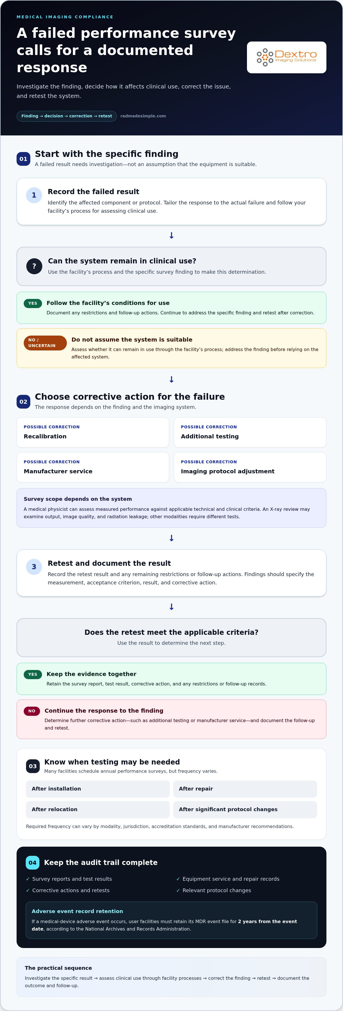
A failed performance survey is a prompt to correct and retest the system, not a finding to file away. Corrective actions for medical imaging compliance equipment may include recalibration, additional testing, manufacturer service, or adjustments to imaging protocols. Requirements vary by modality and jurisdiction. Surveys may also be required after installation, repair, relocation, or significant protocol changes.
A failed result raises immediate questions about safe operation, documentation, and what to address first. The response depends on the finding and the system, so separate equipment performance issues from broader compliance gaps.
This article explains what a failed survey can mean and how to organize the response. It also covers how standards, HIPAA protections, equipment condition, and DICOM safeguards relate to compliance, helping you assess next steps.
Key Takeaways
- Treat a failed performance survey as a finding to investigate, document, correct, and retest before relying on the affected system.
- Separate equipment-performance issues from privacy risks, and assess how access controls and secure data handling protect patient information.
- Identify the standards that apply to each modality and jurisdiction, since requirements can differ across systems and locations.
- Use equipment age, functionality, service history, and compatibility to guide maintenance and replacement planning.
- Strengthen image-data resilience by aligning DICOM safeguards, archiving practices, and recovery planning with operational needs.
What happens if equipment fails a performance survey?
A failed survey identifies a performance result that needs investigation. Record the finding, identify the affected component or protocol, and follow your facility’s process for determining whether the system can remain in clinical use. Tailor the response to the specific failure rather than assuming the equipment remains suitable.
Corrective actions may include recalibration, additional testing, manufacturer service, or an adjustment to imaging protocols. After the correction, document the retest result and any restrictions or follow-up actions. If an adverse event involving a medical device occurs, user facilities must retain its MDR event file for 2 years from the event date, according to the National Archives and Records Administration.
How often must medical imaging equipment be inspected, tested, and serviced? Many facilities schedule annual performance surveys, but the required frequency varies by modality, jurisdiction, accreditation standards, and manufacturer recommendations. Testing may also be needed after installation, repair, relocation, or significant protocol changes.
A medical physicist can assess whether measured performance meets applicable technical and clinical criteria. The survey’s scope depends on the system: an X-ray review may examine output, image quality, and radiation leakage, while other modalities require different tests. Findings should specify the measurement, acceptance criterion, result, and any corrective action.
For onsite testing, arrange access to the imaging room and coordinate with radiology, facilities, and safety staff. Specialized or large X-ray systems are often tested at the facility, where shielding, protective equipment, and radiation-monitoring procedures can be used. After delivery and installation, inspect and test the system, and promptly raise problems with the vendor.
What documentation and records are required for medical imaging compliance audits? Keep survey reports, equipment service and repair records, test results, corrective actions, and relevant protocol changes together. For an adverse event, retain the required MDR event file for the specified period.
How much does medical imaging compliance equipment and testing cost? Cost depends on the case and is provided after a quote request. Modality, testing scope, travel or onsite needs, and corrective work all affect the cost.
What are the risks and penalties for noncompliant medical imaging equipment? Unresolved failures can affect image quality, radiation exposure, patient care, and audit readiness. Regulatory or accreditation consequences depend on the applicable requirements and the nature of the failure.
Use compliance testing resources carefully: FDA regulations are mandatory, while FDA guidance documents provide recommendations. Identify the requirements that apply to the modality and jurisdiction, then keep evidence showing how each finding was addressed.
HIPAA and protected health information
In the United States, HIPAA compliance for imaging systems means protecting patient information wherever the workflow creates, stores, displays, or transmits it. A practical review traces the data from acquisition through diagnostic review, reporting, archiving, and eventual equipment disposal.
HIPAA and Patient Data Protection
The HIPAA Privacy Rule requires covered entities, including owners or operators of medical imaging equipment, to protect individually identifiable health information. Images and associated files can reveal a patient’s identity and care, even when an image appears to have been removed from the system.
Build safeguards around actual access. Use role-based permissions so staff can view only the information needed for their duties, and multi-factor authentication to strengthen account access. Configure automatic logoff on diagnostic workstations, then review audit logs for unusual access or activity.
Protect electronic protected health information both in transit and at rest. Encryption can reduce exposure if data is intercepted or stored on a lost or retired device. Document the risk analysis, mitigation plans, workforce training records, and confidentiality agreements that support your facility’s HIPAA program.
Include every data location in the review. Imaging equipment may store patient information separately from image files, and machines may retain images or associated information after visible images are removed. Before equipment leaves the facility for secondary-market sale, delete protected health information and account for separate storage locations.
FDA and HIPAA Compliance
FDA and HIPAA address different compliance concerns: FDA requirements relate to medical devices, while HIPAA protects patient information in covered healthcare workflows. Understanding both helps facilities assess system suitability without treating device compliance as proof of data privacy.
The FDA states that regulations are mandatory, while guidance documents are recommendations and are not mandatory. When purchasing imaging systems, familiarize yourself with applicable FDA guidance and HIPAA requirements, then assess how the proposed workflow handles patient data.
During procurement and configuration, map where information enters, who can access it, and how it moves between equipment, workstations, and archives. For example, verify that staff access is controlled and image transfers use safeguards appropriate to the facility’s environment. Record decisions in risk analyses and system procedures.
Keep operational controls aligned as protocols, users, or connected systems change. A new diagnostic workflow can affect which staff see patient information and where files are stored. After a workflow change, review permissions, training, audit logging, and encryption, and preserve records of the safeguards in use.
The Health Insurance Portability and Accountability Act of 1996 protects the privacy of patient data in the United States. That responsibility should remain visible as images pass between acquisition, review, reporting, and archiving. Staff should know which permissions apply to their role, where protected information may remain, and how to raise concerns about unexpected access. Before a system is transferred or retired, confirm that the cleanup process covers both image files and associated information, not just what appears on screen. Keeping these checks within routine workflow helps align access, storage, transfer, and disposal practices with the privacy safeguards described above.
Applicable particular standards by equipment type
In the United States, there isn’t one universal equipment checklist for every imaging service. Compliance depends on modality, system components, facility type, and applicable federal and state requirements. Selecting medical imaging compliance equipment starts with identifying the standards that apply to each system and its intended use.
State requirements and testing schedules can vary by modality and jurisdiction. A CT scanner, fluoroscopy system, and mammography unit therefore require different evaluations. Accreditation requirements can also shape a facility’s compliance program.
For U.S. X-ray equipment, manufacturers must comply with applicable Electronic Product Radiation Control requirements in Title 21 CFR Parts 1000 through 1005. Radiation-safety performance standards appear in Title 21 CFR Parts 1010 and 1020. The specific provisions depend on the system’s design and use.
- Diagnostic X-ray systems and major components: 21 CFR 1020.30 sets out applicable requirements.
- Radiographic equipment: 21 CFR 1020.31 addresses this equipment type.
- Fluoroscopic equipment: 21 CFR 1020.32 contains the applicable performance standards.
- Computed tomography equipment: 21 CFR 1020.33 covers CT-specific requirements.
These identifiers help teams match documentation and testing to the system rather than applying one generic checklist. Hospitals also have Medicare Conditions of Participation: 42 CFR 482.26 addresses radiologic services, while 42 CFR 482.53 addresses nuclear medicine services.
Mammography has a distinct framework. Under the Mammography Quality Standards Act, the FDA regulates personnel qualifications, facility quality control and quality assurance programs, and facility accreditation and certification. A mammography review should consider these elements alongside equipment performance.
International standards provide another reference point, but they don’t replace jurisdiction-specific requirements. IEC 60601-1 is the base standard for medical electrical equipment safety and essential performance. IEC 60601-1-3 focuses on radiation protection in diagnostic X-ray systems; IEC 60601-2-43 applies to interventional X-ray equipment, and IEC 60601-2-54 covers radiography and fluoroscopy systems.
For medical imaging compliance equipment, evaluate each modality against its applicable requirements and document how your facility addresses them. Evidence can include performance results, quality assurance records, system documentation, and corrective-action records. Distinguish standards recommendations from binding regulations: FDA regulations are mandatory, while FDA guidance documents are recommendations, not mandatory requirements.
For facilities operating across jurisdictions, compare applicable requirements before transferring a configuration or protocol. A setup accepted in one market may need additional evaluation or documentation elsewhere. This comparison helps align equipment configuration, image workflows, and compliance records with the rules that apply at each site.
Assessing equipment needs and condition
Assess each imaging asset in light of its clinical role, physical condition, service history, and applicable regulatory requirements. A CT scanner used for routine diagnostic work may need a different maintenance plan from ultrasound equipment or a portable system.
Start with an asset-by-asset review. Record the modality, age, functionality, service history, and known performance issues. Compare those details with the manufacturer’s maintenance guidance and relevant safety regulations. This gives you a practical basis for deciding whether to maintain, repair, or plan replacement, rather than relying on age alone.
- Condition: Note recurring faults, component wear, downtime, and changes in image quality.
- Service history: Review maintenance records, repairs, and unresolved recommendations for each system.
- Operational fit: Compare current capacity and functionality with the facility’s imaging needs.
- Compatibility: Assess whether equipment works with existing systems and software.
- Compliance: Identify applicable state regulations and manufacturer requirements for each asset.
Maintenance intervals depend on equipment type. Ultrasound equipment may have daily operator checks and annual preventive-maintenance service. A CT scanner may require daily, quarterly, and annual service intervals. These examples show why one schedule cannot be applied consistently across every modality. Follow the manufacturer’s guidance and relevant safety regulations for each asset when setting its maintenance plan.
Look beyond the system’s exterior. During maintenance, a technologist may disassemble equipment to clean its interior and exterior, then check components and detectors. For a CT scanner, documenting detector condition alongside service findings helps connect observed issues with the system’s performance history. Record the work performed and any follow-up needed in the asset’s maintenance record.
Compatibility affects more than image display. Before adding or replacing equipment, assess its connections with existing systems and software, and involve IT and compliance teams early. For example, a new imaging unit may require workflow changes or configuration work to integrate with the facility’s environment. Identifying these requirements during planning helps clarify the project’s technical and regulatory scope.
Room layout also shapes the decision. Assess the space, patient access, operator access, and applicable state regulations before selecting or relocating equipment. If space is limited, portable medical imaging equipment may help address the constraint, but the room and workflow still need evaluation.
Finally, weigh repair history against current functionality and planned use. Uncertified X-ray components can require additional testing and investigation, increasing project complexity and cost. Include component status in replacement planning, and document why the selected equipment meets operational needs and applicable requirements.
DICOM security, archiving, and disaster recovery
Medical imaging systems and PACS handle identifiable clinical information, so compliance must cover data movement, storage, access, and recovery. Healthcare facilities should protect patient information. Facilities can support that goal with documented security controls and tested recovery procedures.
For DICOM image flows, use TLS where supported and validate both calling and called AE identifiers. Maintain DICOM conformance statements, AE Titles, and modality worklist configurations as system records. These details help teams understand device connections, identify unexpected endpoints, and diagnose a failed transfer without confusing one modality’s configuration with another.
Access safeguards should follow the facility’s risk analysis. Role-based permissions, multi-factor authentication, automatic logoff, and audit logging help control access to electronic protected health information. Encrypt that information in transit and at rest. For example, a facility can review access logs after an unexpected user views a fluoroscopy study, then document its response.
Archiving procedures should define retention schedules, backup frequency, integrity checks, and disaster-recovery RTO/RPO targets. These targets describe recovery expectations for systems and data. Preserve original report content, and use timestamps and electronic signatures to manage addenda and corrections. This creates a traceable clinical record when a radiology report changes after its initial release.
Hospitals must maintain radiology records for at least 5 years, according to the Office of the Federal Register. A retention schedule should align archive handling with that requirement and the facility’s other applicable obligations. Imaging equipment may store operating software on the same hard drives as patient data, so data-clearing procedures must protect both. Removing images alone may leave other patient information behind.
Backups need evidence of recoverability, not just successful completion messages. Imaging centers can test data restores quarterly and document the results. A useful record identifies the archive tested, restore outcome, any integrity issue, and corrective action. Include PACS, modality connections, and clinical access needs in recovery planning so restored data can support the facility’s essential workflows.
- Secure transfers: Configure TLS where supported and validate DICOM AE identifiers.
- Controlled records: Maintain conformance statements, AE Titles, and modality worklist settings.
- Resilient archives: Define backups, integrity checks, retention, and recovery targets.
- Tested restoration: Perform and document quarterly restore tests.
Review your DICOM security and recovery procedures against these safeguards, then explore radiology workflow solutions that support connected imaging environments.
A compliant healthcare imaging provider should use secure data transmission, protect stored imaging results, and control access to patient information. Consider these safeguards together: transmission controls address data in motion, storage protections address archived results, and access controls help limit who can view patient information. Keeping all three in the security program gives staff a practical framework for reviewing imaging workflows and identifying where safeguards need attention.
The article states that future revisions of the IEC 60601 series are expected to place greater emphasis on connected-device security than current editions do. Imaging providers can account for this direction by considering connected-device security during planning and review, while continuing to apply the protections required for their current systems. A documented approach can help teams assess how device connections, image handling, and access controls fit within the facility’s wider security practices.
Dextro Imaging Solutions: Radcom Compact
Dextro Imaging Solutions offers Radcom Compact for facilities assessing their radiology technology needs after a compliance review. For changes to reading workflows or imaging infrastructure, its offerings also include Dextro Reading Stations, PACS Harmony, and aycan PACS. These options can inform an assessment of system requirements, compatibility, and operational priorities.
Use survey findings, equipment condition, and workflow requirements to guide the discussion. For example, a facility reviewing its reading environment can consider Dextro Reading Stations, while a PACS review can include PACS Harmony or aycan PACS. Focus the conversation on the specific operational need rather than treating every compliance issue as a reason to replace equipment.
To discuss your imaging technology needs, contact Dextro Imaging Solutions or visit radmadesimple.com.
Build compliance into your next imaging decision
Make compliance part of equipment planning, not a task reserved for survey preparation. As clinical workflows change, use each purchasing or upgrade decision to consider how the system fits your facility’s technical environment and supports consistent operations. Selecting medical imaging compliance equipment with those needs in view can connect practical workflow priorities with your broader compliance program.
Dextro Imaging Solutions offers Radcom Compact and Dextro Reading Stations. These products offer options to consider when reviewing radiology technology and reading workflows. Identify the operational challenge your team wants to address, then discuss which solution may fit that need.
Contact Dextro Imaging Solutions to discuss your imaging technology needs. A clear plan can help your team move forward with greater focus and confidence.
Frequently Asked Questions
What FDA regulations apply to medical imaging equipment?
FDA requirements for U.S. imaging devices include Electronic Product Radiation Control provisions in Title 21 CFR Parts 1000 through 1005 and performance standards in Parts 1010 and 1020. The applicable provisions depend on modality, such as 21 CFR 1020.33 for computed tomography equipment. Medical Device Reporting regulations in 21 CFR Part 803 also apply to U.S. manufacturers, distributors, importers, and device user facilities. FDA regulations are mandatory; guidance documents are recommendations.
What accreditation standards apply to imaging equipment and facilities, such as ACR and Joint Commission?
Under Section 1834(e) of the U.S. Social Security Act, as amended by the Medicare Improvements for Patients and Providers Act of 2008, qualifying freestanding advanced diagnostic imaging facilities had to obtain accreditation by January 1, 2012, to seek Medicare reimbursement. The three recognized organizations are the American College of Radiology (ACR), the Intersocietal Accreditation Commission, and The Joint Commission. Hospitals also follow Medicare Conditions of Participation for radiologic and nuclear medicine services.
What are the risks and penalties for noncompliant medical imaging equipment?
Unresolved failures can affect image quality, radiation exposure, patient care, and audit readiness. Regulatory or accreditation consequences depend on the applicable requirements and the nature of the failure.


