

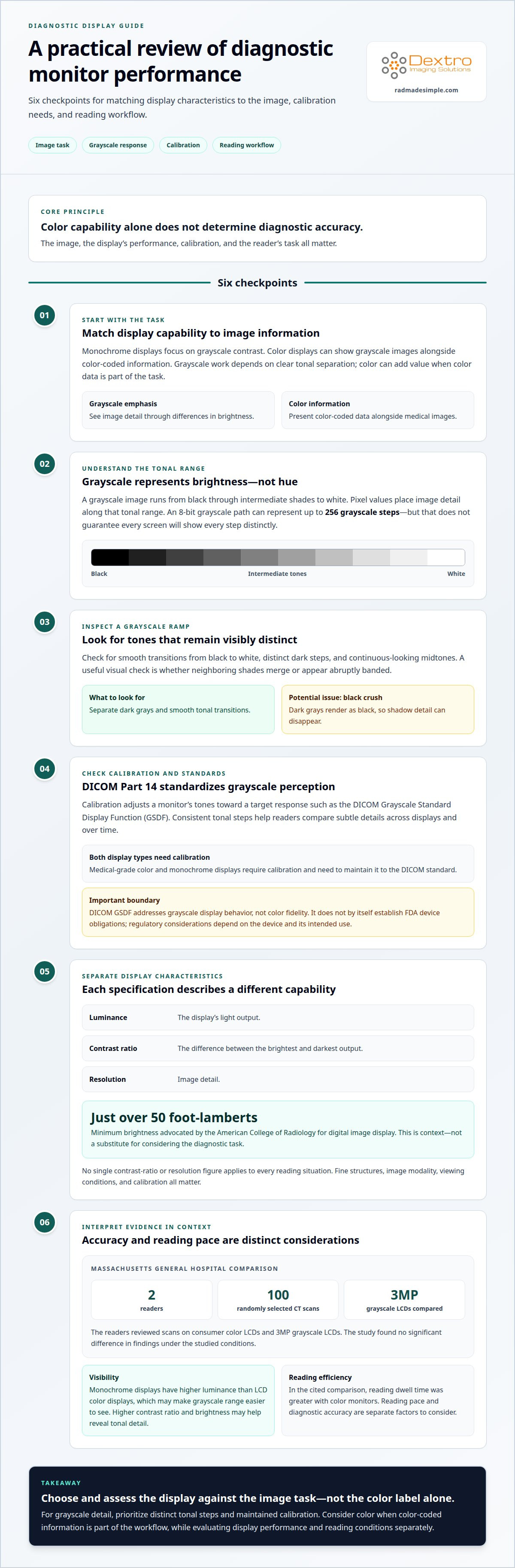
A color display isn’t automatically more accurate for radiography. For clinicians weighing grayscale vs color diagnostic monitors, the key difference is how each presents image information: monochrome displays focus on grayscale contrast, while color displays can show grayscale images alongside color data. DICOM Part 14 standardizes grayscale perception, not color fidelity.
It’s understandable to focus on whether a monitor is grayscale or color, but display type alone does not determine diagnostic accuracy. Calibration, image characteristics, and reading workflow matter, too. This guide to grayscale vs color diagnostic monitors explains how DICOM standards apply, what accuracy findings indicate, and how monitor choice can affect radiology reading efficiency.
You’ll find a practical framework for matching display capabilities to the task, including when grayscale performance is central and when color information adds value. Use this comparison of grayscale vs color diagnostic monitors to identify the factors that matter for your imaging workflow, rather than treating either display type as the universal answer.
Key Takeaways
- Match the display to the image task: grayscale work depends on clear tonal separation, while color can help present color-coded information alongside medical images.
- Understand what grayscale levels describe, and why the display’s ability to distinguish subtle shades matters when reviewing image detail.
- Look beyond monitor type when considering diagnostic accuracy; reading efficiency and the demands of radiography workflows also influence the comparison.
- Explore how Jusha Diagnostic Monitors and Dextro Reading Stations can support a diagnostic reading setup aligned with your imaging workflow.
Grayscale calibration and DICOM standards
For monochrome diagnostic images, viewing depends on consistent grayscale rendering, not simply whether a display is labeled medical-grade. DICOM Part 14 provides the Grayscale Standard Display Function (GSDF) used to standardize grayscale perception.
Grayscale Calibration
Grayscale calibration adjusts a diagnostic monitor so its displayed tones follow a target response, such as DICOM GSDF. Consistent tonal steps help readers compare subtle image details across displays and over time. Both medical-grade color and monochrome displays require calibration and need to maintain it to the DICOM standard. A grayscale ramp can reveal whether dark and light tones remain distinguishable.
DICOM GSDF and US Regulatory Context
DICOM Part 14 is a technical standard for grayscale display behavior, not a specification of color fidelity. It helps make grayscale perception consistent, but it does not define how accurately a monitor reproduces color. For US regulatory questions, distinguish this display-function standard from FDA requirements: DICOM GSDF alone does not establish FDA device obligations. The applicable regulatory considerations depend on the device and its intended use, so the standard should not be treated as a substitute for regulatory review.
Recommended Display Characteristics
Luminance describes the light output of a display, while contrast ratio describes the difference between its brightest and darkest output; resolution describes image detail. The American College of Radiology advocated a minimum brightness of just over 50 foot-lamberts for digital image display. That figure provides context, but luminance, contrast, and resolution serve different purposes and should be considered against the diagnostic task.
No single contrast-ratio or resolution figure applies to every reading situation. A display’s ability to show fine structures and maintain grayscale separation matters alongside the image modality and viewing conditions. For example, reviewing a detailed radiograph places different demands on visible image detail than viewing a worklist. Calibration helps preserve the intended grayscale response at the monitor’s normal operating settings.
Definition of grayscale
Grayscale describes an image using brightness values rather than color hues. A grayscale runs from black through intermediate shades to white, allowing structures to appear through differences in lightness. An 8-bit grayscale path can represent up to 256 grayscale steps. Each pixel’s value determines where it falls along that tonal range, so a small value change can affect how a boundary or texture appears.
Consider a chest image displayed in grayscale: bone, soft tissue, and air appear as different tones, not different colors. The display’s brightness affects how those tones appear, while the image data supplies the pixel values. Picture a ramp from pure black to pure white, with gray steps between them. The more clearly those steps remain distinct, the easier it is to perceive subtle tonal changes.
A scale may be described as containing 256 tones from white to black. In practical terms, that describes the available gradations, not a promise that every screen will show each one distinctly. Some screens render dark grays as black, a problem called black crush. Shadow detail can then disappear into the background. For example, a faint border near a dark region may be harder to distinguish if neighboring shades merge visually.
Grayscale is different from color imaging, which adds hue and saturation information. Color can help present color-coded data, while grayscale represents image detail through luminance changes. Color images are best viewed using Gamma 2.2. For a grayscale image, the display’s tonal response is central to how brightness differences appear. This distinction helps explain why monitor comparisons must account for the image being viewed, not just the screen’s color capability.
To assess the concept in a practical setting, inspect a grayscale ramp and look for smooth transitions from black to white. Notice whether dark steps remain separate and whether midtones look continuous rather than abruptly banded. These observations clarify what grayscale capability means before comparing display technologies. For imaging teams evaluating diagnostic monitor options, the image’s tonal needs provide a useful starting point.
Diagnostic accuracy and reading efficiency
Monitor type can affect image visibility and reading pace, but color alone does not establish diagnostic accuracy. Evidence comparing color and monochrome displays helps distinguish these factors.
Diagnostic Accuracy
Diagnostic accuracy is the ability to identify relevant findings from displayed images; monitor performance can support visibility but cannot guarantee a diagnosis. In a Massachusetts General Hospital study, two readers reviewed 100 randomly selected CT scans on consumer color LCDs and 3MP grayscale LCDs, with no significant difference in findings. This comparison suggests that color displays can support diagnostic reading under studied conditions, rather than proving all displays perform equally.
Monochrome displays have higher luminance than LCD color displays, which can make the grayscale range easier to see. Their higher contrast ratio and brightness may help reveal detail in tonal transitions. The practical comparison is not simply color versus grayscale: image type, display performance, and the reader’s task all matter.
Reading Efficiency
Reading efficiency describes how smoothly a radiologist can review images and reach an interpretation. In the cited comparison, reading dwell time was greater with color monitors, although the study found no significant difference in diagnostic accuracy. A longer review does not by itself mean a less accurate result, but it can matter in a demanding reading workflow.
For example, someone reviewing a sequence of grayscale CT images may value luminance and contrast that make subtle details easier to inspect. A color display can also show color-coded information alongside grayscale images. The more useful choice depends on whether that additional color information supports the task or adds little to the reading process.
Ambient Lighting and Viewing Conditions
Ambient lighting changes how a monitor’s brightness and contrast appear. Reflections or a bright room can make dark image regions harder to distinguish, while a dim room can make shadow detail easier to assess. Consider the viewing environment alongside the monitor’s normal operating brightness, rather than treating it as a separate factor.
To assess shadow detail, use a dim room and set monitor brightness near its normal daily level, rather than maximum. This shows whether dark tones remain visible under familiar viewing conditions. A display that looks clear only at maximum brightness may look different during routine reading.
Radiography diagnostic reading comparison
Radiologists generally use grayscale displays when interpretation relies on subtle tonal detail, and color displays when color information supports the task. Color monitors can display mammography, CT, MRI, and ultrasound images, but suitability depends on the display’s performance for the intended reading task.
Choosing Grayscale or Color Monitors
Choosing between grayscale and color means matching display capabilities to the image and reading task. For radiographs with subtle positive findings, luminance and visible tonal detail are important considerations. A Massachusetts General Hospital comparison selected 100 radiographs with subtle positive findings and added 30 normal radiographs, focusing the evaluation on challenging cases.
That comparison helps explain why color capability alone doesn’t establish an advantage for grayscale interpretation. Early color CRT monitors could not show the minimum 256 shades of gray distinctly enough for diagnostic radiological images. In comparing grayscale vs color diagnostic monitors, consider whether the display makes subtle tones clear for the specific radiographs being read.
Monitor Suitability Across Imaging Types
Suitability for mammography, CT, MRI, and ultrasound depends on the image task and the display’s ability to present its detail. A color monitor can show grayscale images, but color capability alone does not establish suitability for diagnostic reading. The useful comparison is whether grayscale information remains clear and whether any color data supports the task.
For CT, a Massachusetts General Hospital study compared consumer color LCDs with 3MP grayscale LCDs. Two readers reviewed 100 randomly selected CT scans, with no significant difference in findings. That result applies to the tested CT comparison, not automatically to mammography, MRI, or ultrasound. The practical choice between grayscale vs color diagnostic monitors should account for the modality and reading needs.
The grayscale LCDs in the radiography comparison had 3-megapixel resolution and ran at 150 foot-lamberts. These are characteristics of the displays in that study, not universal specifications for every modality. Backlight and display characteristics can affect how tones appear, so compare displays using the image type and detail readers need to assess.
For an imaging workflow that includes radiography, explore reading workstation options suited to diagnostic image review.
Dextro Imaging Solutions: Jusha Diagnostic Monitors
Dextro Imaging Solutions offers advanced diagnostic displays for imaging teams choosing monitors for diagnostic reading. They are a relevant option when your workflow calls for medical imaging monitors and you’re comparing grayscale performance, color capability, and reading needs.
The right setup involves more than the display. Dextro Reading Stations and PACS Harmony are also available for teams considering their reading environment and imaging workflow alongside monitor selection. The choice depends on the images being reviewed and how the workstation fits into daily radiology operations.
If you’re assessing display options for your imaging workflow, contact Dextro Imaging Solutions to discuss your needs.
Make Your Next Display Decision With Confidence
Choosing between grayscale and color diagnostic monitors is best treated as a workflow decision, not a matter of choosing the most advanced-looking screen. Consider the image types your team reads, the details that matter in those images, and how the display fits into the reading environment. That context can turn technical comparisons into a practical direction for your department.
Dextro Imaging Solutions offers Radcom Compact, Dextro Reading Stations, and Jusha Diagnostic Monitors. These options help imaging teams consider monitor and workstation needs together, rather than treating the display as an isolated purchase. The next step is to discuss how your imaging workflow shapes those requirements.
Contact Dextro Imaging Solutions to discuss your diagnostic monitor needs. A clear understanding of your workflow is a strong starting point for choosing a setup that supports your team.
Frequently Asked Questions
How Are They Different?
The two types differ in how they present image information: grayscale displays focus on intensity changes, while color displays can also show hues. When comparing two images side by side, grayscale presentation can keep attention on tonal differences; color may be useful when an image includes color-coded information. The better fit depends on what the reader needs to see and how the images are used.
How Are They Similar?
Both types can display medical images, and both medical-grade color and monochrome displays require calibration. A color display can show grayscale images, while a monochrome display focuses on shades rather than color hues. For example, a department using color for worklists and grayscale imaging for review still needs its diagnostic display to render image tones consistently.
LED Displays and Grayscale: Why Is It Important to Know the Relationship?
LED describes a display’s illumination technology, while grayscale describes how image tones range from dark to light. The terms refer to different aspects of a screen, so an LED label alone does not tell you how clearly it renders grayscale detail. A grayscale test ramp, for example, can help reveal whether adjacent dark tones appear distinct on the display.
What Is A Monochrome Display?
A monochrome display presents images using a single color channel, typically as shades of gray from black to white. A color display can present multiple hues. For example, a monochrome screen may show a radiograph through tonal variation, while a color screen can also present color-coded information in an image or interface.
What Is the Difference Between Grayscale and Color Diagnostic Monitors?
The difference lies in how each display handles image information: grayscale monitors focus on tonal variation, while color monitors reproduce hues as well. This can shape how a reader views an image and supporting information on screen. A radiology team comparing the options should consider whether its work relies chiefly on grayscale image detail or also uses color-coded data during review.
What Display Standards Apply to Diagnostic Monitors, Such as DICOM GSDF and AAPM TG18?
DICOM Part 14 defines the Grayscale Standard Display Function, or GSDF, for standardized grayscale perception. AAPM TG18 provides display assessment guidance and test patterns used to evaluate image presentation. They serve related but different roles: DICOM GSDF describes a grayscale response, while TG18 supports evaluation of display performance. Neither term by itself describes color fidelity.


