

The most future-ready radiology department isn’t the one that adopts every new technology first. Future-proofing a radiology department means planning for change with adaptable spaces, connected imaging and information systems, and AI that supports radiologists rather than replacing their judgment. The goal is to respond more easily as equipment, workflows, and patient needs evolve.
That takes more than choosing new technology. Space planning, interoperability, and practical workflow decisions all affect how well a department can adapt without adding unnecessary complexity. AI can help prioritize urgent findings, but radiologists remain central to interpreting images in their clinical context.
This article explains how to plan flexible imaging spaces, connect systems, and use AI as a supportive tool. It also covers how reading stations, PACS, and voice solutions can contribute to a more adaptable radiology workflow.
Key Takeaways
- Plan imaging spaces with future equipment changes in mind, so upgrades can fit without requiring a full redesign.
- Connect imaging, reporting, and communication workflows to help teams share information across systems; health information systems can use HL7 and DICOM standards for reliable data sharing, according to the U.S. Government Publishing Office.
- Use AI to help flag and prioritize urgent findings while keeping radiologists central to image interpretation and clinical decisions.
- Explore flexible workstation and PACS options to support changing reading needs and more adaptable workflows.
1. Flexible space for future imaging equipment upgrades
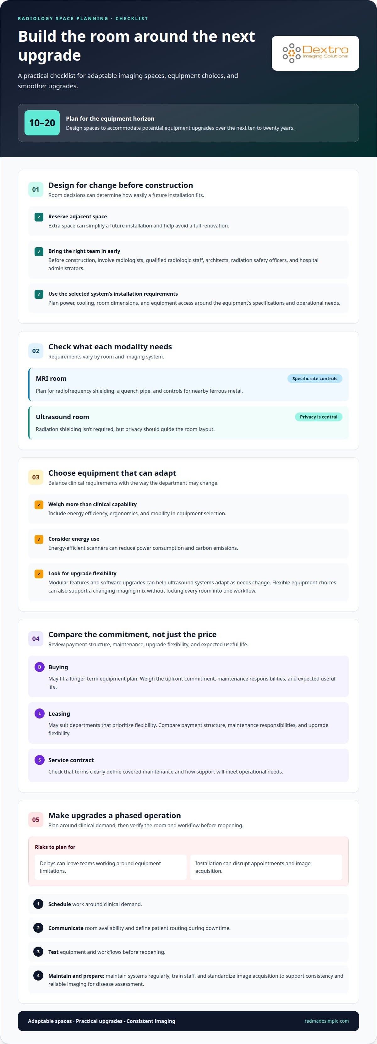
Planning for future-proofing a radiology department starts with the room, not just the scanner. Design spaces to accommodate potential equipment upgrades over the next ten to twenty years. Reserving adjacent space can simplify future installation and help avoid a full renovation. Before construction begins, involve radiologists, qualified radiologic staff, architects, radiation safety officers, and hospital administrators.
Facility requirements vary by modality. An MRI room needs radiofrequency shielding, a quench pipe, and controls for nearby ferrous metal. Ultrasound rooms don’t require radiation shielding, but privacy remains central to the layout. Use the selected system’s installation requirements to plan power, cooling, room dimensions, and equipment access. Working with power protection specialists such as Energy Control Systems can also protect sensitive diagnostic hardware against electrical disturbances and voltage anomalies. This helps ensure the room meets operational needs.
When selecting equipment, weigh energy efficiency, ergonomics, and mobility alongside clinical requirements. Energy-efficient scanners can reduce power consumption and carbon emissions. Modular features and software upgrades can help ultrasound systems adapt as needs change. Flexible equipment choices can also accommodate a changing imaging mix without locking every room into one workflow.
Compare buying, leasing, and service-contract options by weighing upfront commitments, payment structure, maintenance responsibilities, upgrade flexibility, and expected useful life. Leasing may suit departments that prioritize flexibility, while buying may fit a longer-term equipment plan. Service-contract terms should clearly define covered maintenance and how support meets operational needs.
In practice, future-proofing a radiology department also means managing upgrade risks. Delays can leave teams working around equipment limitations, while installation may disrupt appointments and image acquisition. Plan changes in phases: schedule work around clinical demand, communicate room availability, and define patient routing during downtime. Before reopening, test equipment and workflows. Maintain systems regularly, train staff, and standardize image acquisition to support consistency and reliable imaging for disease assessment.
2. Adapting to technology, costs, globalization, and patient knowledge
Healthcare technology and patient expectations keep changing how radiology teams deliver care. The Centers for Disease Control and Prevention notes that imaging studies increase by up to 5% annually. Departments can adapt by reviewing referral patterns, reducing duplicate examinations, and matching imaging capacity to clinical need. Global collaboration and secure virtual access can also help colleagues share expertise across locations while keeping patient care central.
Radiologists add value by interpreting images in light of a patient’s anatomy, physiology, disease process, and clinical question. Access to clinical colleagues supports appropriate examination choices and discussion of complex findings. A centralized department layout can keep imaging services together and reduce patient movement. Clear signs, accessible routes, private changing areas, and a calm waiting space support comfort, privacy, and wayfinding.
Make onboarding efficient before the appointment. Collect complete patient information upfront, send preparation instructions in advance, and use automated messages for reminders and preparation steps. Clear communication about accessing images and reports can also help patients. These steps reduce avoidable confusion without replacing direct communication when clinical questions arise.
For U.S. modernization, assess applicable radiation-safety requirements, accreditation criteria, and payer or reimbursement policies for each proposed modality and workflow. Relevant considerations depend on the service and care setting. There’s no single upgrade cost: budget needs depend on equipment, facility changes, system integration, training, maintenance, and implementation; equipment pricing is established after a quote request.
Workforce planning should pair emerging-technology training with protected time to practice new workflows. The Association of American Medical Colleges projected that the shortage of radiologists and other specialists could exceed 35,000 by 2034, while radiology residency positions grow by about 2% annually. Track performance through measures such as reporting turnaround, equipment downtime, repeat imaging, and patient flow. Compare these indicators before and after changes to assess operational value and identify where further adjustment is needed.
3. Integration with EMR, PACS, and AI platforms
Connect imaging equipment, the radiology information system (RIS), picture archiving and communication system (PACS), and electronic medical record (EMR) around a clear workflow. The RIS manages scheduling and exam information; PACS stores and displays images; the EMR gives physicians access to reports and patient records. Health information systems can use HL7 and DICOM standards to support reliable data sharing across settings, according to the U.S. Government Publishing Office; the stated value is 7.
Start by mapping how an order moves from referral to completed report. Confirm that patient identifiers and examination details transfer consistently from the EMR or RIS to imaging equipment and PACS. Then test whether images and reports return to the correct patient record. A sample workflow should cover an order, image transfer, radiologist review, and report delivery. This helps teams identify interface gaps before a wider rollout.
For PACS and image storage, plan capacity around image volume, retention needs, access patterns, and recovery processes. Evaluate cloud or hybrid storage alongside on-premises infrastructure, considering network performance, data access controls, backups, and how staff retrieve prior images. Radiologists comparing current and previous studies need a consistent viewing workflow, whether images are stored locally or remotely.
Cybersecurity planning should cover user access, system updates, backups, and incident response across connected platforms. Define who can access images, reports, and administrative functions, then review permissions as roles change. Before integrating AI analytics, identify where results appear in the reading workflow and how radiologists review them. Test software interfaces with representative cases, document failures, and establish a process for maintaining integrations after upgrades. These steps support reliable performance without disrupting reporting or adding avoidable work.
Choose systems that integrate with existing EMR, PACS, and AI analytics platforms. Interoperability solutions can help radiology departments coordinate imaging, reporting, and communication systems. Radiology reports can be transmitted securely to referring healthcare providers through electronic medical record systems.
4. AI as a support for radiologists
AI adds value when it helps radiologists prioritize work, not when it replaces clinical judgment. In a hospital workflow, an algorithm can flag suspected acute findings so a radiologist can review urgent studies sooner. The radiologist still interprets images in context, considering the patient’s clinical question, history, and other findings. This keeps AI in a supporting role.
Start with a defined use case. For example, a department might introduce triage software to identify studies with suspected critical findings. Map where alerts appear, who reviews them, and how the team handles an alert that differs from the initial assessment. Test the process with representative cases before using it in routine reporting. Clear roles help ensure an AI prompt does not replace review.
Research cited on the page says AI can reduce image-reading turnaround time by about 60%. Treat that as potential, not an automatic result: actual workflow impact depends on the tool, its integration, and how staff use it. Measure reporting time, alert volume, and radiologist workload before and after implementation. Also review whether the tool enhances prioritization without creating unnecessary interruptions.
AI capabilities can include flagging acute abnormalities and supporting stroke-outcome prediction. These functions address different clinical tasks, so each needs a defined workflow and review process. Radiologists should understand what the system flags and where its output appears alongside images. Regular case reviews and team feedback can help show whether the technology supports diagnostic decisions and patient outcomes.
Choose tools that fit existing image-review routines, then train staff using realistic examples. Quality image display remains part of that workflow, alongside software and AI. Explore Dextro Imaging Solutions as an option for the diagnostic image-review environment.
Dextro Imaging Solutions: Portable Radiology Workstations
Dextro Imaging Solutions offers Portable Radiology Workstations for departments adapting reading workflows across locations. They can be useful when teams need workstation options as they plan flexible access to imaging and reporting. The right setup depends on the department’s workflow and existing systems.
For a broader reading environment, Dextro Reading Stations and Radcom Compact are also available. PACS Harmony can be part of planning how image access and PACS workflows fit into department operations. These offerings support different parts of a radiology technology setup, so the right fit depends on the department’s needs.
If your department is evaluating workstation or PACS options as its needs change, contact Dextro Imaging Solutions to discuss your requirements.
Turn planning into a practical next step
Future-proofing a radiology department is an ongoing planning discipline, not a one-time technology purchase. A useful next step is to identify the workflow constraint that most affects your team, then align future decisions around it. That might mean improving how radiologists access studies, supporting consistent image review, or accommodating changing clinical demands.
Technology choices should work together within a clear operational plan. Dextro Imaging Solutions offers Radcom Compact, Dextro Reading Stations, and Jusha Diagnostic Monitors for departments planning their reading and diagnostic environments. Choose options according to the workflow needs your team has prioritized.
With a focused requirement in hand, you can move from broad modernization goals to a more specific conversation about suitable tools. Contact Dextro Imaging Solutions to discuss your department’s needs. Take the next step with a plan that keeps radiologists and patient care at its center.
Frequently Asked Questions
What is the Future of Radiology?
The future of radiology is a more connected, data-informed service where imaging, reporting, and clinical decisions work together. Progress should mean appropriate examinations, accessible results, and clear communication for patients, not simply newer scanners. Departments can assess each technology against a defined care need, its effect on daily workflows, staff capability, and its fit with existing systems.
What does future-proofing a radiology department involve?
Future-proofing involves making technology and facility decisions that keep options open as clinical needs evolve. For example, when planning an imaging-room refresh, document which workflows must continue during the work, assign ownership for system updates, and set criteria for reviewing future equipment needs. This turns modernization into an ongoing operational plan rather than a series of disconnected purchases.
Which imaging technologies and modalities should radiology departments invest in for the next 5-10 years?
There is no single modality mix that suits every department. Prioritize imaging capabilities that match local referral needs, patient populations, and clinical services. A service with growing ultrasound demand may prioritize adaptable ultrasound capacity, while another may focus on maintaining its MRI pathway. Before committing, compare each investment’s clinical use, staff readiness, facility fit, and compatibility with existing workflows.
How should radiology departments plan for AI, automation, and clinical decision support?
Start with one clearly defined task, such as prioritizing studies flagged for possible urgent findings. Set a review process that shows who receives the alert, how it is assessed, and how disagreements are documented. Involve radiologists and clinical colleagues in testing representative cases, then monitor whether the tool supports decisions without creating distracting alerts. Keep professional interpretation and accountability central to patient care.


