

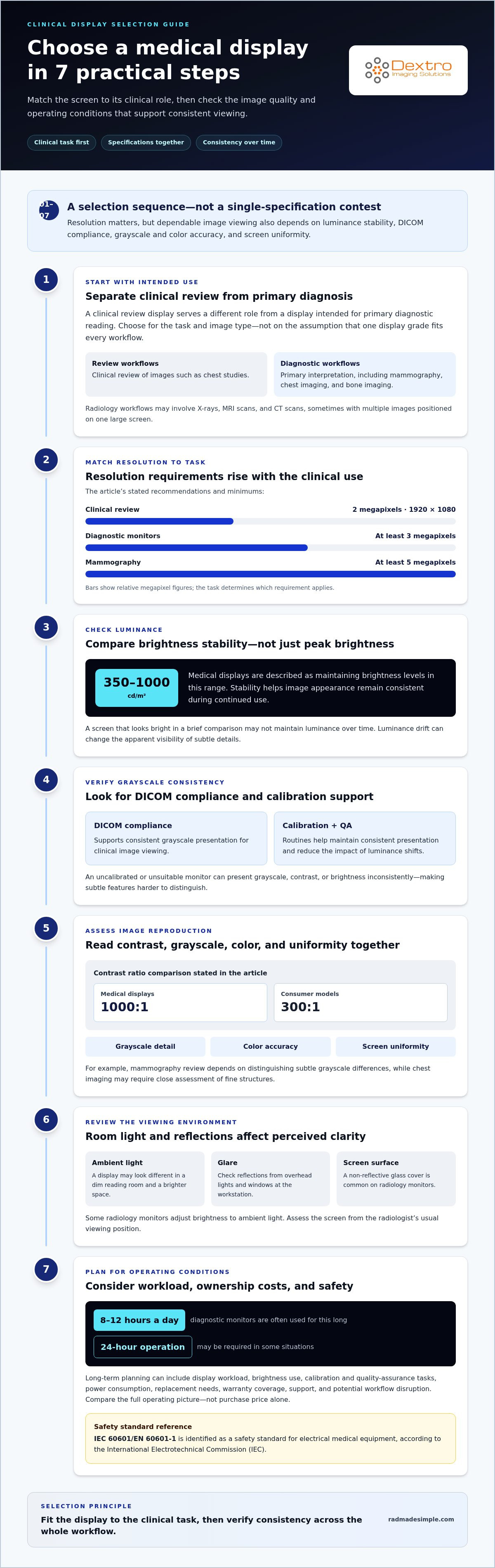
The highest-resolution monitor isn’t automatically the best fit for every clinical review. When comparing medical imaging displays, start with the task: a clinical review display is commonly recommended at 2 megapixels, or 1920 × 1080 pixels, while mammography calls for at least 5 megapixels. The right choice depends on the images clinicians need to assess and whether the display is used for review or primary diagnosis.
Resolution matters, but it’s only one part of dependable image viewing. Brightness stability, DICOM compliance, grayscale and color accuracy, and screen uniformity all affect how consistently image details appear. A display can perform well in one area yet fall short if luminance shifts over time or subtle shades are difficult to distinguish.
This guide explains how these specifications work together and what they mean in practice. You’ll learn how to compare resolution and pixel pitch, evaluate grayscale and color reproduction, and understand why calibration and brightness consistency matter. Use this framework to match medical displays to clinical workflows without treating any single specification as the whole answer.
Key Takeaways
- Match display resolution to the clinical task: clinical review and primary diagnostic reading can call for different display requirements.
- Compare brightness stability alongside peak brightness, since consistent luminance supports reliable viewing over time.
- Look for DICOM compliance and calibration features that help maintain consistent grayscale presentation.
- Assess contrast, grayscale and color accuracy, and screen uniformity together to understand how clearly image details may appear.
- IEC 60601/EN 60601-1 is identified as a safety standard for electrical medical equipment, according to the International Electrotechnical Commission (IEC).
Medical Display Guide: How to Select Medical Monitors for Clinical Reviews?
Choose a monitor based on its clinical role, image type, and workflow. A display for reviewing chest images has different demands from one used for mammography or primary diagnosis.
Diagnostic Performance Across Display Grades
Diagnostic performance describes how reliably a display presents image details for a particular clinical task. It matters because radiologists and doctors use those details to assess findings, compare studies, and guide decisions. Medical-grade displays are designed for diagnostic reading, while consumer displays may suit less complex review tasks. Match the display to its intended use rather than assuming one grade fits every workflow.
Radiology workflows use displays for X-rays, MRI scans, and CT scans, sometimes positioning multiple images on one large screen. Mammography, chest imaging, and bone imaging are examples where medical-grade display technology should remain the standard. For primary reads, use a display suited to diagnostic interpretation; clinical review monitors serve a different role.
Medical Display Uses and Specifications
A medical imaging display is a monitor used to present clinical images for review or diagnosis. Comparing medical imaging displays means looking at resolution, luminance, contrast, and intended specialty together. Clinical review displays are commonly recommended at 2 megapixels, or 1920 × 1080 pixels. Diagnostic monitors are described as needing at least 3 megapixels, while mammography requires at least 5 megapixels.
Medical displays are described as maintaining brightness levels of 350-1000 cd/m², and a 1000:1 contrast ratio is compared with 300:1 for consumer models. IEC 60601/EN 60601-1 is identified as a safety standard for electrical medical equipment, according to the International Electrotechnical Commission (IEC). These specifications help frame selection, but the specialty and reading purpose determine which requirements matter most.
Lifespan and Ownership Costs
Lifespan and total cost of ownership include more than the monitor’s purchase price. Display workload, brightness use, calibration, quality-assurance tasks, power consumption, and replacement needs all affect long-term planning. Diagnostic monitors are often used 8 to 12 hours a day, and some situations require 24-hour operation, so operating conditions matter.
Typical prices for medical imaging displays in the United States depend on display grade, resolution, calibration features, and included support; pricing is provided after a quote request. Warranty terms also shape ownership costs by determining which repairs or replacements are covered and for how long. Compare the full operating picture, including calibration and potential workflow disruption, rather than considering purchase cost alone.
Risks of Unsuitable Displays
An unsuitable or uncalibrated monitor can present grayscale, contrast, or brightness inconsistently. That may make subtle image features harder to distinguish, especially when a radiologist compares studies on different screens. A consumer display used for a primary diagnostic read may not provide the image presentation that task requires.
For example, mammography review depends on distinguishing subtle grayscale differences, while chest imaging may require close assessment of fine structures. Luminance drift can also make an image appear different over time. Calibration and quality-assurance routines help maintain consistent presentation, supporting dependable radiology workflows without making resolution the only selection criterion.
Medical and consumer display brightness stability
Brightness stability affects whether radiology images retain a consistent visual appearance over time. Compare it alongside ambient viewing conditions and screen-wide uniformity, rather than considering maximum brightness in isolation.
Brightness Stability and Image Clarity
Brightness stability is a display’s ability to maintain luminance during continued use, helping images remain visually consistent. Medical displays are described as maintaining brightness levels of 350-1000 cd/m² for years. That stability matters when radiologists review images across reading sessions, because luminance changes can alter the apparent visibility of subtle details.
A consumer screen may look bright during a brief comparison, but peak brightness alone doesn’t show how consistently it will present images over time. Compare displays using the same image and viewing conditions, then consider how brightness maintenance supports their intended applications. For example, subtle structures in a chest image can be harder to assess when the image looks dimmer or brighter than expected.
Brightness, Glare, and Viewing Conditions
Viewing conditions shape how brightness translates into image clarity because room light and screen reflections can affect visual perception. A display that looks clear in a dim reading room may be harder to assess in a brighter space. Some radiology monitors adjust brightness to ambient light, helping adapt the screen to changing room conditions.
Glare can obscure details or distract the viewer, so assess reflections from overhead lights and windows at the workstation. A non-reflective glass cover is common on radiology monitors. Compare the display from the radiologist’s usual viewing position rather than relying only on a straight-on product image. Consistent room lighting also helps clinicians compare studies without environmental changes affecting their visual impression.
Brightness controls should support a stable viewing setup, not simply make every image look brighter. Increasing screen brightness can’t restore image information that isn’t present in the source. Instead, match luminance to the room and keep the viewing setup consistent when assessing image quality.
Uniformity and Brightness Enhancement
Brightness uniformity describes how evenly luminance appears across the screen. It matters because a bright center and dim edge can make the same image look different depending on its location. Medical-grade displays may use uniformity correction to improve consistency across the screen. This is useful when images span large areas or clinicians compare multiple views side by side.
Calibration and quality-assurance software can help maintain stable, DICOM-compliant images over time. These tools support reliable grayscale presentation, but they don’t replace suitable display hardware. When comparing displays, assess the center and edges using the same image, and note whether contrast and brightness appear consistent across the viewing area.
For a more complete assessment, compare displays in their intended radiology workflow and consider radiology workflow guidance alongside brightness specifications. This keeps the decision focused on image viewing, room conditions, and the practical needs of clinical applications.
Image quality, luminance stability, and DICOM compliance
Image quality depends on grayscale reproduction, contrast, color accuracy, luminance uniformity, and stability. When selecting displays in the United States, distinguish image-presentation calibration from electrical safety standards and facility accreditation requirements.
DICOM Compatibility and Calibration
DICOM Part 14 compliant calibration supports consistent grayscale reproduction across medical monitors. A display following the DICOM grayscale standard display function, or GSDF, presents luminance steps in a controlled relationship. This helps radiologists compare image details consistently at a workstation, including when they review studies from different devices.
DICOM compatibility is also a workflow consideration. The monitor, workstation, and imaging software need to handle medical image data in a way that preserves its intended presentation. For example, a mammography image viewed on a workstation should retain its grayscale relationships rather than being altered by unrelated display settings. DICOM compliance alone doesn’t establish that every display suits every diagnostic task.
For U.S. practice, the identified standards include DICOM Part 14 calibration for grayscale presentation and IEC 60601/EN 60601-1, identified as a safety standard for electrical medical equipment, according to the International Electrotechnical Commission (IEC). These standards address different considerations; neither statement alone establishes every facility’s accreditation requirements.
Luminance Stability and Quality Assurance
Luminance stability describes how consistently a monitor maintains its brightness over time. A panel’s output can change with use, which may affect how image tones appear during patient studies. A comparison simulated display luminance values for panels that had been in use for one year, illustrating why assessment should consider performance beyond initial setup.
A practical quality-assurance framework can include regular calibration, testing, and performance verification. Automated quality-assurance and calibration software can help maintain stable images while minimizing workflow interruptions. These checks give imaging teams a repeatable way to detect drift and keep a medical monitor aligned with its intended grayscale presentation.
Calibration supports reliable image viewing, but it doesn’t replace appropriate display selection. A mammography workstation, for example, must be evaluated for the imaging task as well as its calibration process. Quality assurance connects device performance to everyday use, helping teams identify changes before they become routine viewing conditions.
Image Quality in Clinical Use
Image quality combines several characteristics, not brightness alone. Contrast affects separation between adjacent tones, while grayscale accuracy supports visibility of subtle differences. Color accuracy matters when images include color-coded information. Uniformity helps keep luminance consistent across the screen, so an image doesn’t appear different near its center and edges.
For a patient study, these factors shape what the radiologist can visually assess. Compare medical monitors using representative images and the intended viewing workflow. Then review calibration records and quality-assurance results together. This approach connects technical performance to actual image presentation, rather than treating a compatibility label as a complete measure of diagnostic suitability.
Contrast, grayscale, color accuracy, and uniformity
These image-quality characteristics work together: contrast separates tones, grayscale preserves subtle differences, and uniformity keeps luminance consistent across the screen.
Contrast and Low-Contrast Structures
Contrast describes the difference between bright and dark areas, helping reveal structures with similar surrounding tones. Medical displays are compared at a 1000:1 contrast ratio, versus 300:1 for consumer models. The distinction matters when radiologists assess low-contrast features, where limited separation can make subtle structures harder to distinguish from adjacent tissue.
A ratio alone doesn’t describe every aspect of image quality. The source image, display settings, and viewing conditions also affect how clearly low-contrast structures appear. Compare the same clinical image on each screen, paying attention to faint borders and transitions instead of judging contrast from a bright demonstration image.
Grayscale, Color Accuracy, and Uniformity
Grayscale accuracy describes how faithfully a monitor presents shades between black and white, while color accuracy concerns how closely displayed colors match intended values. Medical displays are described as having color deviation below ΔE 2, compared with above ΔE 5 in consumer models. These differences matter when image interpretation relies on subtle shades or color-coded details.
Medical displays with 10-bit or higher grayscale can show 1024 or more shades, compared with 256 on consumer models. Mammography may require distinguishing between six similar shades of white to detect microcalcifications. Uniformity correction helps maintain more consistent brightness and color across the screen, so a feature doesn’t appear different near an edge.
These characteristics depend on the full image path, not just the panel. A medical graphics card or display controller processes image data before it reaches the monitor; the workstation and display settings also shape the result. Compare the same image and settings across displays, then assess grayscale transitions, colors, and luminance from center to edge.
- Contrast: Check whether adjacent tones remain distinguishable in low-contrast images, rather than relying on a ratio alone.
- Grayscale: Consider the displayed tonal range, especially for tasks such as mammography, where similar white shades matter.
- Color: Compare color accuracy when clinical images use colored information; ΔE indicates color deviation.
- Uniformity: Look for consistent brightness and color across the screen, including corners and edges.
- Signal chain: Evaluate the monitor with its graphics card, display controller, workstation, and image settings in the intended workflow.
Assessing these features together gives a more useful comparison than selecting by a single specification. A display with strong contrast can still produce uneven luminance, while grayscale depth alone doesn’t establish color accuracy. For a practical evaluation, use representative clinical images and examine subtle tonal transitions as well as screen-wide consistency.
What specifications matter when comparing medical imaging displays?
Start with the clinical task, then compare resolution, grayscale depth, viewing angle, pixel pitch, and calibration support. These specifications work together; a strong result in one category can’t compensate for a mismatch elsewhere.
Resolution sets how much image detail a screen can represent. Clinical review displays are recommended at 2 megapixels, or 1920 × 1080 pixels. Diagnostic monitors are described as needing at least 3 megapixels, while mammography requires at least 5 megapixels. Match the monitor to its intended use rather than treating these categories as interchangeable.
Pixel pitch also influences how tightly pixels sit together. For clinical review, the recommended pixel pitch is 0.250 millimeters and should not exceed 0.300 millimeters. Consider it alongside screen size and resolution: the combination affects how image details appear from the normal workstation position. For example, a large screen can help position multiple images together with fewer manipulations.
Grayscale depth and viewing angle add important context. A medical display with 10-bit or higher grayscale can show 1024 or more shades, compared with 256 on consumer models. A wide viewing angle of at least 178° can reduce substantial head and eye movements when reading radiology images. These specifications matter in shared workstations or layouts where clinicians view the screen from different positions.
- Resolution: Match the display to the use case, such as 2 megapixels for clinical review or at least 5 megapixels for mammography.
- Pixel pitch: Compare the 0.250-millimeter clinical review recommendation with the 0.300-millimeter maximum.
- Grayscale: Assess whether 10-bit or higher output supports the tonal detail needed for the images reviewed.
- Viewing angle: Consider at least 178° when workstation placement or shared viewing affects posture.
- Calibration and uniformity: Include DICOM support and screen-wide consistency in the evaluation, not just panel resolution.
Use the same representative images and workstation settings when comparing medical imaging displays. Check whether fine structures remain visible, grayscale steps stay distinct, and the screen remains consistent from center to edges. Then weigh those observations against the specialty, reading workflow, and number of images viewed together. This turns a specification sheet into a practical selection framework, without assuming one monitor suits every clinical application.
Dextro Imaging Solutions: Radcom Compact
Dextro Imaging Solutions offers Radcom Compact for teams evaluating equipment for medical imaging workflows. Once display requirements are clear, match the monitor and workstation to how clinicians review images and manage their daily workflow.
For a display-focused evaluation, specialized diagnostic monitors are a relevant option. Dextro Reading Stations and Portable Radiology Workstations provide workstation choices, while PACS Harmony supports imaging workflow needs. These offerings give radiology departments and imaging centers distinct categories to consider alongside display specifications.
Use the criteria in this guide to clarify your clinical tasks and priorities, then explore the options that fit your setup. Contact Dextro Imaging Solutions to discuss your imaging equipment needs.
Turn Display Criteria Into a Confident Next Step
Make display selection part of a broader imaging-workflow decision. Before procurement, align clinical users and technical teams on how each monitor will be used, where it will sit, and how it fits the workstation environment. A shared plan helps departments evaluate equipment consistently and supports a smoother transition into daily use.
For teams assessing their setup, Dextro Imaging Solutions offers Radcom Compact, Dextro Reading Stations, and Jusha Diagnostic Monitors. These options provide a starting point for matching display and workstation needs to clinical workflows. The goal of comparing medical imaging displays is not simply to select a screen, but to choose an approach that fits image review and department operations.
Explore the available options and discuss your imaging equipment needs with Dextro Imaging Solutions. Contact Dextro Imaging Solutions to take the next step with a clearer plan.
With the right criteria in place, your team can move forward with focus and confidence.
Frequently Asked Questions
What is a diagnostic display?
A diagnostic display is a medical monitor used to view images for primary interpretation, such as X-rays, MRI scans, and CT scans. Its role goes beyond showing an image for general reference: radiologists assess visible features to interpret a study. For example, a reading workflow may place related images side by side, allowing the radiologist to compare views while evaluating the same examination.
What are the differences between diagnostic and clinical review displays?
A diagnostic display supports primary image interpretation, while a clinical review display supports viewing images outside that primary-reading role. This distinction affects which display specifications suit the task. For example, a clinician reviewing a study during a consultation may need a different setup from a radiologist completing a primary read. Identify who will view the images and how they’ll use them before choosing a display category.
How do medical displays compare with consumer monitors for diagnostic imaging?
Medical displays are designed for clinical image presentation, while consumer monitors serve general viewing purposes. Their brightness stability, grayscale handling, and calibration capabilities can differ. Elizabeth A. Krupinski said consumer-grade displays could be used for less complex imaging classes such as ultrasound, MRI, and CT. That doesn’t make every consumer monitor suitable for every task; intended use and diagnostic role remain central to selection.
What are the DICOM GSDF and DICOM Part 14 calibration requirements?
DICOM Part 14 defines a grayscale presentation function, and GSDF refers to the Grayscale Standard Display Function used to calibrate luminance steps. A quality-assurance schedule can help teams monitor calibration and display performance over time. In practice, teams can document calibration and test results by workstation, making it easier to track display performance and identify changes over time.
How do grayscale and color medical displays compare for different imaging tasks?
Grayscale displays emphasize tonal differences in images such as X-rays and mammograms, where subtle shades can carry important visual information. Color displays support images that encode information through color, including pathology applications. For primary reading with color displays, medical-grade monitors are recommended. Match the display to the image type and interpretation task, then assess whether grayscale detail or accurate color presentation matters most in that workflow.


