

Choosing a mammography interpretation display involves more than resolution, and the appropriate specifications depend on the imaging workflow. Digital mammography monitor specifications therefore need to be evaluated in context: FFDM and DBT workflows affect what the display must support for image review. Resolution matters, but it isn’t the only consideration.
It’s understandable to look for one number that settles the choice. The 5MP benchmark is useful, but a sound evaluation also considers calibration, viewing conditions, and the requirements of the imaging workflow.
This article explains how FFDM and DBT shape display evaluation, what to include in a review document, and how grayscale calibration, room lighting, and 5MP monitors fit into the decision. You’ll have a clearer framework for assessing a diagnostic display without treating a single specification as the whole answer.
Key Takeaways
- Assess display needs in the context of FFDM and DBT workflows, not resolution alone.
- Use a structured review document to capture image-review requirements and relevant U.S. mammography facility obligations.
- Understand how DICOM grayscale presentation and hardware calibration support consistent image display.
- Consider room lighting, screen reflections, and the role of 5MP monitors together when evaluating image-review conditions.
What should the mammography monitor review document?
A useful evaluation document shows how a display supports the intended breast-imaging workflow. Connect technical measurements with image presentation, viewing modes, and applicable U.S. requirements.
Display Performance Measures
Document the display’s resolution, luminance, grayscale reproduction, and calibration, along with each test method and result. This lets reviewers assess whether image detail is presented consistently. Grayscale performance matters because breast imaging relies on subtle differences across an image, while calibration helps maintain the intended grayscale presentation over time.
Poor calibration or display quality can make subtle details harder to distinguish and affect interpretation. The cost of a medical-grade mammography monitor depends on factors such as resolution, screen size, calibration features, and system configuration. Request a quote for pricing.
Viewing Modes and Screen Format
Viewing modes and screen format describe how a display presents images for different review tasks. Document whether multi-resolution, focus, and pathology modes are available, and how each changes image presentation. Focus mode, for example, may support closer examination of a selected region, while multi-resolution viewing can help readers move between detail and overview.
Record screen size and aspect ratio alongside the viewing modes. These factors affect how images fit on screen and how readers arrange multiple views. Distinguish the display’s physical dimensions from its image resolution, since they describe different properties.
Pixel Detail for Breast Imaging
Pixel detail includes both the image’s pixel resolution and the display’s pixel dimensions. Record both, along with the intended imaging workflow. Pixel dimensions indicate how many pixels form the displayed image; resolution describes the level of detail the display can present.
For breast imaging, assess whether the display preserves relevant detail at the viewing scale used for interpretation. Note any scaling or image interpolation, since changing an image’s displayed size can affect how pixel detail appears. A clear record helps distinguish detail in the source image from the display’s capabilities.
FDA Requirements for Diagnostic Monitors
In the United States, MQSA regulations in 21 CFR Part 900 set requirements for covered mammography facilities, including certification, accreditation, image quality, and quality assurance, according to the U.S. Government Publishing Office.
MQSA does not universally require every interpretation monitor to have FDA clearance specifically for FFDM or DBT. The ACR strongly recommends FDA-cleared monitors for mammography interpretation, while noting that other devices may legally be used. Record the monitor’s intended use and clearance status to distinguish facility requirements from display-selection guidance and give reviewers a clear basis for evaluating the system.
DICOM compliance and hardware calibration
DICOM Part 14 defines the Grayscale Standard Display Function (GSDF), a framework for presenting grayscale values consistently. Calibration aligns a monitor’s output with that function, helping preserve tonal distinctions as images move through a clinical interpretation workflow. DICOM compliance alone doesn’t establish that a display is suitable for every mammography task; hardware performance and quality control also matter.
Record whether the display is calibrated to GSDF, how luminance is assessed, and when calibration and testing take place. Keep the results with the monitor’s identification and intended clinical use. This creates a technical record for tracking changes over time and distinguishes a configured display from one evaluated for diagnostic use.
A qualified medical physicist must test diagnostic review workstations before clinical use and regularly thereafter. The ACR strongly recommends FDA-cleared monitors for mammography interpretation. The ACR also states that the FDA recommends monitors and printers specifically cleared for FFDM use by its Office of Device Evaluation, although other devices may legally be used.
MQSA sets quality-assurance requirements for covered mammography facilities, but it doesn’t universally require every interpretation monitor to have FDA clearance specifically for FFDM or DBT. In practice, the ACR’s recommendation and the facility’s quality-control process should inform monitor evaluation. The 2018 ACR Digital Mammography Quality Control Manual is an FDA-approved alternative to the manufacturer’s QC manual.
What are the ACR and MQSA recommendations for mammography monitor quality control and testing? Have a qualified medical physicist test diagnostic review workstations before clinical use and regularly afterward. Follow facility quality assurance procedures, and consider the ACR’s recommendation for FDA-cleared monitors alongside the FDA’s stated clearance guidance.
Document the calibration method, luminance results, test dates, and corrective actions together. That record helps connect hardware changes with image interpretation conditions. A structured mammography display workflow can make these technical checks easier to track.
Screen reflection and ambient lighting
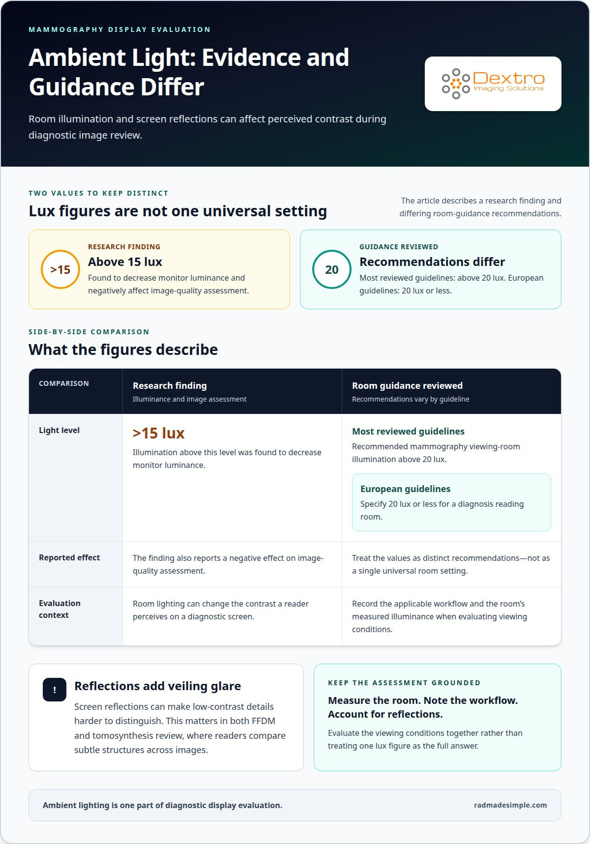
Room lighting changes the contrast a reader perceives on a diagnostic screen. In mammography viewing conditions, illumination above 15 lux was found to decrease monitor luminance and negatively affect image-quality assessment. Reflections add veiling glare, which can make low-contrast details harder to distinguish. These effects matter for both FFDM and tomosynthesis review, where readers compare subtle structures across images.
Room guidance differs. Most guideline documents reviewed recommended mammography viewing-room illumination above 20 lux, while European guidelines specify 20 lux or less for a diagnosis reading room. Treat these values as distinct recommendations, not a single universal setting. The relevant evaluation should record the applicable workflow and the room’s measured illuminance.
Separate diagnostic reading conditions from image acquisition conditions. A 2021 audit in breast screening units in North West England reported mean reporting-room illuminance of 12.32 ± 4.6 lux. The same audit reported mean acquisition-room illuminance of 105.3 ± 178.8 lux. These different readings show why a single room-light target shouldn’t be applied indiscriminately across both workflows.
Record ambient illuminance at the reading position, noting nearby light sources, window reflections, and fixtures facing the screen. Check for reflections on the active image area, then document the room layout and lighting conditions. Repeat the assessment under routine interpretation conditions rather than relying on a measurement taken in a different setup.
Where possible, control glare at its source. Adjusting fixtures or reducing direct light on the screen can limit reflections without changing the displayed image. After adjusting the lighting, reassess contrast, especially when reviewing dense breast tissue or comparing multiple views. A consistent room setup supports dependable viewing and helps distinguish changes in image appearance from changes in room conditions.
5-Megapixel mammography monitors
A 5MP display is a common reference point in mammography evaluation, but resolution alone can’t establish diagnostic suitability. The practical takeaway is to assess pixel count alongside the workstation, intended workflow, and quality assurance.
Five-Megapixel Monitor Characteristics
A 5-megapixel monitor has five million display pixels, providing a defined resolution benchmark for reviewing mammography images. A 5MP display is commonly cited as an industry minimum for mammography interpretation, but the FDA doesn’t mandate a specific monitor resolution.
The benchmark gives teams a useful starting point for comparing displays, not a complete acceptance test. Record the monitor’s resolution and intended use in the workstation evaluation, then consider those details alongside image presentation and the facility’s quality-control process. The review found that monitors with 3 or 5 MP seemed preferred, showing that selection involves more than pursuing the largest pixel count.
Diagnostic Comparison by Monitor Resolution
An 8MP display offers a different pixel count from a 5MP monitor, but that difference doesn’t by itself demonstrate improved diagnostic performance. In a University of Arizona study, radiologists spent an average of 10% less time viewing and scanning mammography images on a single 8MP screen than on dual 5MP monitors.
This finding describes viewing time, not diagnostic accuracy or clinical outcomes. Compare configurations by considering how readers view images, how the workstation supports their workflow, and how quality assurance evaluates the complete setup. A single higher-resolution screen and a dual-monitor arrangement offer different layouts; pixel count alone does not determine which is appropriate.
For field digital mammography, use the resolution figure as one documented selection criterion. Pair it with the workstation’s intended clinical role and a defined quality-control process. Calibration and ongoing evaluation help establish whether the display continues to perform as expected; they don’t make resolution a substitute for the rest of the assessment. If monitor selection is part of a broader workstation decision, explore mammography workstation options as part of that review.
Dextro Imaging Solutions
Dextro Imaging Solutions helps imaging teams align diagnostic displays and reading workflows with their review requirements. High-grade diagnostic monitors are an option to consider when an evaluation identifies a need for mammography-focused display hardware, alongside the facility’s calibration and quality-control processes.
For a broader workstation decision, consider Dextro Reading Stations or Radcom Compact alongside PACS Harmony for imaging workflow. The right fit depends on how the facility reviews FFDM and DBT images, organizes diagnostic work, and maintains display quality. These products connect monitor selection with the workstation and image-management setup, rather than treating resolution as the only consideration.
Contact Dextro Imaging Solutions to discuss the display and workflow requirements for your evaluation.
Turn display requirements into a clear next step
Make your display evaluation useful beyond the purchasing decision. Turn the criteria for digital mammography monitor specifications into a practical plan for assessing, maintaining, and reviewing the workstation as clinical needs evolve. Assign responsibility for quality-control records, define how changes to the workstation or reading environment will be assessed, and keep the rationale for the selected configuration accessible to the team.
This connects technical decisions with day-to-day interpretation and gives your team a clear reference when workflows change, equipment is updated, or review procedures need refinement. A considered plan supports consistent decisions without reducing monitor suitability to a single feature.
If you’re planning a display or workstation review, contact Dextro Imaging Solutions to discuss your requirements. A focused conversation can help you identify the relevant considerations for your imaging environment and determine a practical direction for the next stage. Move forward with a plan that supports your team’s work.
Frequently Asked Questions
How do FFDM and DBT affect display evaluation?
Evaluate FFDM and DBT against the images and review tasks used in each workflow. FFDM review focuses on presenting mammography images, while DBT review involves navigating tomosynthesis images. Assess how the workstation presents and moves through each image type, including whether readers can maintain an overview while examining specific details. The display setup should support the clinical review process in practice.
What information should procurement gather before selecting a display?
For procurement, document the intended clinical use, image types, workstation arrangement, and quality-control process. Specify whether the display will support FFDM, DBT, or both, and whether readers use one screen or multiple screens. Include the planned calibration approach, room conditions, and how image presentation fits existing systems. This information helps procurement compare options against operational needs, not just product labels.
Which display characteristics should a facility assess?
Assess resolution, luminance, grayscale presentation, calibration, screen format, and reflections under interpretation conditions. Record the display model, intended use, measured performance, and test date together. Consider how the display handles image placement and whether readers can inspect relevant views without disruptive rearrangement. Evaluate the complete workstation, since connected equipment and viewing conditions also shape the experience.
What are the minimum resolution and pixel pitch requirements for digital mammography monitors?
A 5MP display is commonly cited as an industry minimum for mammography interpretation, but the FDA does not mandate a specific monitor resolution. The digital mammography monitor specifications should also identify pixel pitch, which describes spacing between pixels. Consider pixel pitch alongside resolution and image presentation, rather than treating it as an independent pass-or-fail measure. The practical evaluation is whether the complete display supports the intended image-review workflow.
What luminance and contrast ratio should a diagnostic mammography monitor have?
U.S. monitor requirements do not specify a single luminance or contrast-ratio figure. Evaluate luminance in the calibrated state and under the room conditions used for reading. Since ambient light can affect perceived contrast, document measurement conditions alongside results. This distinguishes display performance from environmental effects and makes later comparisons meaningful when the workstation, room layout, or monitor changes.
What grayscale bit depth and DICOM GSDF calibration are required for mammography displays?
DICOM Part 14 describes the Grayscale Standard Display Function, which supports calibrated grayscale presentation. The available requirements do not establish a universal grayscale bit-depth minimum for mammography displays. Record bit depth as part of the technical evaluation, then assess whether the calibrated system presents grayscale consistently for its clinical use. Keep that information with the calibration and quality-control records for the workstation.


