

Could a 2MP display be the right choice for your diagnostic workflow, or do its headline specifications leave important questions unanswered? The jusha m260g is described as a 21.3-inch monochrome diagnostic monitor with 1600 × 1200 resolution and a maximum brightness of 1000 cd/m². Those figures help define the display, but they don’t establish whether it fits a particular reading task.
Resolution, grayscale processing, calibration, and intended use all affect how a monitor performs in an image-review workflow. Product descriptions may not explain those practical distinctions, and published LUT claims can differ. For a useful assessment, compare the exact model’s current documentation with the task, workstation, and applicable requirements.
This guide explains the M260G’s reported features and how to assess their relevance to your workflow. You’ll learn what to consider when evaluating a 2MP monochrome display, including calibration and quality assurance, intended use, and PACS and workstation integration. The goal is a clear, evidence-based assessment of fit, grounded in the specifications and documentation for your market and use case.
Key Takeaways
- The jusha m260g’s feature labels are a starting point, not proof that it fits a specific diagnostic task.
- Understand how grayscale mapping supports image display, and why bit depth alone doesn’t establish image accuracy.
- Assess monitor suitability against the modality, image type, clinical task, applicable requirements, and workstation setup.
- Use a structured validation process to review model identity, current documentation, intended use, and market-specific requirements.
- Plan monitor selection alongside calibration, PACS access, workstation configuration, installation, and ongoing support.
What Is the Jusha M260G? A Clear Overview of the 2MP Monochrome Display
Available product listings describe the Jusha M260G as a Jusha-branded 2MP monochrome diagnostic display. That identifies its broad category, but not whether it suits a particular imaging task. Radiologists, imaging IT teams, and procurement staff should assess the exact model against its intended use, current manufacturer documentation, and the complete reading workflow.
Three factors are related but distinct: resolution is the pixel count, grayscale capability describes how image tones are represented, and diagnostic performance depends on the display and its operating conditions as a whole. Calibration, workstation configuration, and the images being reviewed all affect the practical result.
What does 2MP monochrome mean for medical image viewing?
Megapixels measure the number of pixels a display can show. A 2MP screen has roughly two million pixels, but that figure alone doesn’t establish how accurately a medical image appears or whether the display suits a clinical task.
Monochrome displays present images in shades of gray, which can support review of grayscale-focused images. Suitability still depends on the modality, image type, clinical task, applicable requirements, and viewing conditions. A source image may contain more pixels than the display can show at once, so the software may scale it or require zooming and panning. Ambient light, display settings, and workstation configuration can also affect what the viewer sees.
Which M260G specifications are reported in available product content?
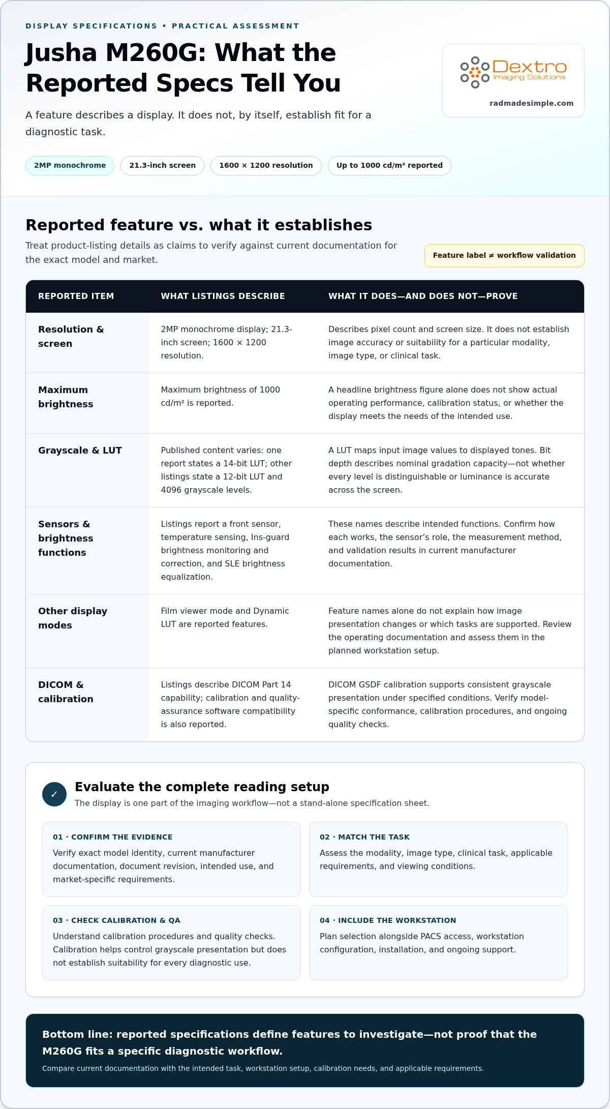
Available product content describes the M260G as a 2MP monochrome display with a 21.3-inch screen and 1600 × 1200 resolution. It also reports a 14-bit look-up table (LUT), a front sensor, temperature sensing, and compatibility with calibration and quality-assurance software. Treat these as reported details, then compare them with the current manufacturer datasheet, intended use, calibration requirements, and market-specific documentation. Published grayscale claims can vary between sources, so confirm the model and document revision before relying on a specification.
A LUT maps input image values to displayed tones. Greater bit depth alone doesn’t prove that a display reproduces images accurately in a clinical workflow. Listings also describe DICOM Part 14 capabilities. The Digital Imaging and Communications in Medicine (DICOM) standard includes guidance for consistent grayscale display, but any model-specific conformance claim should be verified in current manufacturer documentation.
A published specification describes a display feature. It doesn’t prove the display is suitable for a particular diagnostic workflow.
How the M260G’s Grayscale and DICOM Features Are Intended to Work
Feature names describe intended functions, not independently verified image performance. Available product listings report a 12-bit look-up table (LUT), 4096 grayscale levels, Ins-guard, SLE, film viewer mode, and Dynamic LUT for the jusha m260g. Other published material may state different LUT specifications. Confirm each figure and feature against current manufacturer documentation for the exact model and market.
A LUT translates incoming pixel values into display output. In grayscale imaging, that mapping determines how image values appear as shades from dark to light. A stated 12-bit LUT and 4096 possible levels describe nominal gradation capacity. They don’t demonstrate that every level is distinguishable, that luminance is accurate across the screen, or that the display is calibrated for a particular diagnostic task.
What do Ins-guard, SLE, and Dynamic LUT claim to do?
Listings describe Ins-guard as a real-time brightness-monitoring and correction feature, and SLE as a function intended to equalize brightness across the screen. These descriptions explain intended behavior, not the correction’s accuracy or the screen’s uniformity in a working setup. Current manufacturer documentation should explain the exact operation, sensor role, measurement method, and validation results.
Film viewer mode and Dynamic LUT are also reported features. Their names alone don’t establish how they change image presentation or which tasks they support. Review the operating documentation and evaluate the functions within the planned workstation configuration before relying on them in a clinical workflow.
What does DICOM grayscale calibration mean?
DICOM grayscale calibration adjusts a display’s luminance response to follow the DICOM Grayscale Standard Display Function (GSDF), supporting consistent presentation of grayscale steps under specified conditions. Calibration helps control how image values map to visible luminance, but it doesn’t by itself establish suitability for every diagnostic use. Verify the requirements for the intended task and market, including calibration procedures and ongoing quality checks. The FDA guidance for diagnostic display devices offers a reference for evaluating display performance in diagnostic radiology. Its relevance depends on the device’s intended use and market.
For a practical evaluation, look beyond the feature list. Confirm the current specifications, understand how calibration is performed, and assess the display as part of the reading workstation. Explore radiology workflow planning resources to connect display evaluation with the wider imaging setup.
Is a 2MP Monochrome Monitor the Right Fit for Your Imaging Workflow?
A 2MP label describes resolution, but it can’t determine whether a display suits a diagnostic task. Fit depends on what clinicians review, how images are presented, applicable requirements, and how the monitor works with the workstation. Treat the jusha m260g’s published specifications as inputs to an evaluation, not proof of diagnostic performance or regulatory compliance.
Start with the clinical task and modality, then compare the display against those needs. X-ray, CT, and MRI workflows can involve different image types and review practices, so avoid assumptions based on modality or pixel count alone. For broader context on Jusha displays, see the Jusha medical monitor reference guide.
How should radiologists compare display resolution and grayscale needs?
Use a task-based comparison to organize the evaluation. It won’t establish suitability on its own, but it helps teams identify what to verify before deployment.
| Factor | What to assess | Verification question |
|---|---|---|
| Resolution | Pixel dimensions relative to image detail and viewing method | Will images be viewed at native size, scaled, zoomed, or panned? |
| Grayscale | Required tone presentation for the images and review task | What calibration and grayscale performance documentation applies? |
| Clinical task | Image type and the decisions supported by its review | Is the display intended and validated for this use? |
| Modality | X-ray, CT, or MRI images within the actual workflow | Do the image characteristics and workstation setup match the intended use? |
Keep display specifications separate from clinical outcome claims. A resolution or grayscale figure doesn’t establish that a monitor improves diagnostic accuracy or meets requirements for a particular setting.
What should buyers assess beyond the model name?
Assess luminance behavior and screen uniformity, the calibration method and schedule, ambient lighting, and the quality-control process for detecting performance changes. Check that the graphics hardware, operating system, image-viewing software, connections, and PACS workflow support the planned configuration. A monitor’s capabilities matter only when the full reading setup can use and maintain them.
Requirements may differ by clinical task, organization, and market. Review current manufacturer documentation alongside applicable authoritative guidance, and record how the display will be tested and maintained. The 2026 radiology equipment compliance checklist offers broader prompts for organizing that review.
How to Validate M260G Before Adding to a Reading Setup
Move from the clinical task to the documentation, then evaluate the complete workstation. This sequence helps radiologists, imaging IT teams, and procurement staff identify gaps before deployment.
- Define the task. Record the modality, image types, intended reading use, and display configuration the workflow requires.
- Confirm model identity. Match the model name on the unit and documentation. Check that the datasheet applies to that exact model and review its revision date.
- Review intended use and documentation. Compare listed features with current manufacturer materials. Check the intended use, calibration method, quality-control expectations, maintenance requirements, and market-specific regulatory or compliance documentation.
- Map system compatibility. Compare the monitor’s documented inputs and requirements with the workstation, graphics hardware, operating system, image-viewing software, PACS workflow, and number and arrangement of displays.
- Test the configured setup. Evaluate the display with the intended software and image workflow. Confirm that calibration and quality-control procedures can be carried out and documented for the deployment.
Assess product specifications against the intended clinical workflow, rather than treating them as a stand-alone measure of suitability.
Which documents and performance details should a team review?
Keep a record of the datasheet version reviewed and compare each claimed feature with supporting manufacturer documentation. Look for details on calibration, luminance and uniformity measurement, quality-control intervals, and maintenance. Check regulatory and compliance statements against authoritative documentation for the relevant market and intended use. A feature name or general product description does not establish compliance for a specific deployment.
How does the monitor fit into a workstation and PACS workflow?
Map the jusha m260g to the actual reading station, including graphics hardware, display connections, operating-system version, image-viewing software, PACS access, and any additional displays. Compare each component with documented system requirements, then test the configured workflow to identify compatibility or setup issues before clinical use. For system-level planning, see the radiology reading workstation guide.
A coordinated setup brings the monitor, workstation, PACS integration, installation, and ongoing support into one evaluation. Plan your radiology workstation integration around the needs of the complete imaging workflow.
Plan a Jusha Monitor Evaluation Around the Complete Imaging System
A monitor decision is strongest when it starts with the clinical task and considers the whole image-review setup. For the jusha m260g, evaluate intended use alongside current manufacturer specifications, calibration needs, and compatibility with the workstation and PACS workflow. A feature list can inform the decision, but the configured system determines how the display fits into daily work.
What should an imaging team align before deployment?
Bring clinical, IT, and procurement requirements together before implementation. Confirm which image-review tasks the display must support, then align the selection with workstation specifications, PACS access, and the intended display arrangement. This helps teams identify practical dependencies early, such as available connections, software requirements, and calibration procedures.
Plan implementation responsibilities as well. Document who will coordinate installation, complete initial calibration and user setup, carry out quality checks, and manage maintenance. Verify relevant model documentation and requirements for the intended market and workflow. Base decisions on current evidence rather than assuming a feature label establishes suitability, compliance, or a specific clinical outcome.
How can Dextro support a broader monitor and workstation plan?
Dextro Imaging Solutions offers Jusha diagnostic monitors alongside specialized reading stations and portable radiology workstations. Its imaging-system planning also connects workstation configuration with PACS integration, installation, and technical support. Considering these components together helps teams plan around how images are accessed, displayed, and reviewed, rather than treating the monitor as a stand-alone purchase.
As part of a system plan, compare documented display requirements with the workstation and PACS environment, then account for calibration and ongoing support in the deployment process. Dextro’s Jusha monitors, workstations, and imaging workflow solutions can support an evaluation of how equipment and software fit into the broader setup.
For a coordinated view of medical imaging hardware, workstations, and PACS integration, explore Dextro’s medical imaging solutions.
Build a Reading Setup Around Verified Workflow Needs
The jusha m260g’s reported specifications can help frame an evaluation, but resolution and feature names alone don’t establish whether it suits a diagnostic task. Confirm the current manufacturer documentation, intended use, calibration requirements, and compatibility with the workstation and PACS environment before making a decision.
Plan the monitor as part of the full imaging system. Display configuration, installation, quality checks, and ongoing technical support all contribute to a workable reading setup. Dextro offers Jusha diagnostic monitors and specialized radiology workstations, with installation, technical support, and imaging software integration to help connect equipment to the broader workflow.
Ready to plan your imaging environment with the complete system in view? Explore Dextro’s medical imaging solutions and take the next step toward a setup aligned with your team’s needs.
Frequently Asked Questions
What resolution is the Jusha M260G monitor?
Available product information reports a native resolution of 1600 × 1200 pixels for the Jusha M260G. That resolution describes the number of pixels on the display, not whether it can show every source image at its original size or meet a particular reading task’s requirements. Check the current manufacturer datasheet for the exact model and market, then assess resolution alongside image type, viewing method, and workstation configuration.
Is the Jusha M260G a monochrome diagnostic display?
Yes. Product listings describe the M260G as a 2MP monochrome diagnostic display. Monochrome means it presents images in shades of gray, a format used for grayscale-focused image review. The category label alone doesn’t establish suitability for every modality or clinical task. Confirm the monitor’s documented intended use and applicable requirements, then consider how it will be calibrated and configured within the reading workflow.
What does the M260G’s 12-bit LUT mean?
A 12-bit look-up table (LUT) can represent 4096 possible values, which the display system maps to output tones. That figure describes nominal gradation capacity, not proof that all levels are visibly distinct or accurately calibrated. Published M260G descriptions differ on LUT bit depth, so verify the exact claim against a current manufacturer datasheet for the specific model. Bit depth alone doesn’t establish diagnostic performance.
How does the Jusha M260G’s Ins-guard feature work?
Some product listings describe Ins-guard as a feature for real-time brightness monitoring and correction, reportedly using a built-in sensor. That description indicates the claimed purpose, but doesn’t independently validate how the function operates or its performance in a particular setup. Review current manufacturer documentation for its measurement method, correction behavior, calibration relationship, and quality-control requirements before including the feature in a deployment plan.
Can a 2MP monitor be used for diagnostic radiology?
A 2MP monitor may be suitable for some diagnostic workflows, but the resolution label alone can’t determine that. Suitability depends on the modality, image type, clinical task, applicable requirements, calibration, and workstation configuration. Define the intended reading use first, then compare it with the monitor’s current specifications and documentation. Don’t treat a product category or pixel count as proof of regulatory compliance or clinical suitability.
Does the Jusha M260G support DICOM calibration?
Available product information reports DICOM-related grayscale capabilities and compatibility with calibration and quality-assurance software. Review current manufacturer documentation to establish exactly which calibration functions apply to the M260G, how they’re performed, and whether they match the intended use and market. A DICOM-related feature claim by itself doesn’t confirm that a display is calibrated, maintained, or appropriate for a specific diagnostic task.
What should I verify before integrating a diagnostic monitor with PACS?
Check the monitor’s documented inputs and requirements against the workstation, graphics hardware, operating system, image-viewing software, and PACS environment. Confirm the supported display configuration, intended image-review tasks, calibration process, and quality-control responsibilities. Test the complete setup with the planned workflow before deployment. Reviewing these components together helps identify compatibility and implementation gaps that a monitor datasheet or PACS specification alone may not reveal.


