

The highest-resolution screen isn’t automatically the right choice for diagnostic reading. Choosing X-ray diagnostic monitors means weighing image detail alongside grayscale performance, calibration, viewing conditions, and how the display fits your PACS and workstation. Specifications can be hard to compare across display categories, so focus on the features that matter for the clinical task and daily workflow.
Resolution matters, but so do consistent image presentation, quality assurance, and the demands of the imaging work your team performs. This guide explains what to compare when evaluating displays for diagnostic X-ray interpretation, including DICOM grayscale performance, PACS and reading-station integration, and ongoing display quality. The goal is to plan a complete reading environment, not simply choose a monitor with impressive specifications.
Key Takeaways
- Choose X-ray diagnostic monitors around the clinical tasks they’ll support, not resolution alone.
- Compare resolution, pixel pitch, luminance, contrast, and grayscale performance as related factors.
- Match display capabilities to the reading setup, and distinguish diagnostic displays from screens used for general tasks.
- Involve radiologists, imaging leadership, biomedical engineering, and IT in evaluation, deployment, and ongoing quality checks.
- Assess how Jusha diagnostic monitors fit into the complete reading environment, including workstation and PACS requirements.
What Makes a Monitor Suitable for Diagnostic X-Ray Reading?
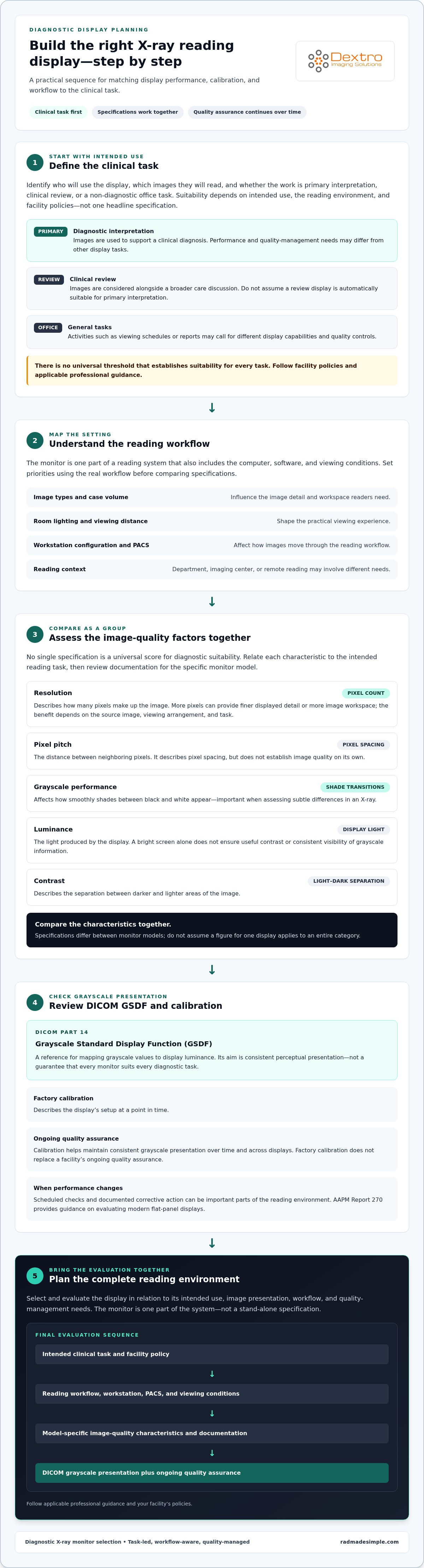
Start by defining the work the display needs to support. A monitor used for primary interpretation may have different performance and quality-management requirements from one used to review images alongside a patient’s record or handle office tasks. The right choice depends on the clinical task, reading environment, and facility policies, not a single headline specification.
A diagnostic monitor is a display selected for a defined medical imaging task, with image presentation and quality management suited to that intended use. This definition puts the focus on the full setup: how the image appears, how display performance is evaluated, and whether the monitor fits the reading workflow. X-rays create images from differences in how tissues absorb radiation, so consistent presentation matters when professionals assess them. See this overview of X-ray technology for foundational context.
Diagnostic interpretation versus clinical review
Primary diagnostic interpretation uses images to support a clinical diagnosis. Clinical review may involve checking images as part of a broader care discussion, while an office display may be used for non-diagnostic tasks such as viewing schedules or reports. These activities can call for different display capabilities and quality controls. Don’t assume that a screen used for review is automatically suitable for primary interpretation, or that a display used for diagnostic reading is the best fit for general office tasks.
There isn’t one universal threshold that establishes suitability for every task. Before selecting a display, identify its intended use and follow facility policies and applicable professional guidance. Requirements can depend on the image type, clinical setting, and how the display will be used.
Why X-ray workflow shapes the display decision
The same monitor may serve different roles in a radiology department, imaging center, or remote reading setup. Image types and case volume influence the amount of workspace and image detail readers need. Room lighting, viewing distance, workstation configuration, and the way images move through PACS also affect the practical experience. Use these factors to set priorities before comparing specifications.
For example, a high-volume reading station may need to accommodate a different application layout and pace of work than a display used for occasional image review. Remote reading introduces additional considerations about the workstation and viewing environment. The monitor is one part of the system, alongside the computer, software, and conditions in which images are viewed. Explore the radiology reading workstation guide for more on how these elements fit together.
When evaluating X-ray diagnostic monitors, first identify who will use the display, which images they’ll read, and whether the task is primary interpretation or review. This gives your team a practical basis for comparing technical characteristics.
Which Image Quality and Calibration Factors Matter for X-Ray Monitors?
Display specifications describe different aspects of an image, but they don’t combine into one universal score for diagnostic suitability. Assess resolution, pixel pitch, luminance, contrast, and grayscale performance together and in relation to the intended reading task. For X-ray diagnostic monitors, the practical question is whether the display presents relevant image detail consistently within the facility’s workflow.
Resolution, grayscale, and luminance in context
Resolution describes how many pixels make up the image. More pixels can provide finer displayed detail or more image workspace, but the benefit depends on the source image, viewing arrangement, and clinical task. Pixel pitch is the distance between neighboring pixels. It describes how closely the pixels are spaced, but it doesn’t establish image quality on its own.
Grayscale performance affects how smoothly shades between black and white appear. This matters when a reader assesses subtle differences in an X-ray. Luminance is the light a display produces, while contrast describes the separation between darker and lighter areas. A bright screen alone won’t ensure useful contrast or consistent visibility of grayscale information.
Compare these characteristics as a group, then check the documentation for the specific monitor model. Specifications can differ between models, so don’t assume a figure for one display applies to an entire category.
DICOM GSDF calibration and quality assurance
DICOM is the DICOM standard for medical imaging, covering the exchange and management of medical images. For display presentation, DICOM Part 14 defines the Grayscale Standard Display Function (GSDF), a reference for mapping grayscale values to display luminance. The aim is consistent perceptual presentation, not a guarantee that every monitor suits every diagnostic task.
Calibration helps maintain consistent grayscale presentation over time and across displays. A manufacturer’s factory calibration describes the display’s setup at a point in time; it doesn’t replace a facility’s ongoing quality assurance. Because display performance can change, scheduled checks and documented corrective action can be important parts of the reading environment. AAPM Report 270 provides guidance on evaluating modern flat-panel displays. Facilities should follow applicable professional guidance and their own policies.
For a practical look at DICOM considerations, read the DICOM monitor compliance guide. Teams planning an integrated reading environment can also explore diagnostic display and workstation options alongside PACS workflow needs.
How Do X-Ray Diagnostic Monitors Compare Across Reading Setups?
Compare displays in the context of the reading environment, rather than assuming there is one ideal screen or monitor count. A dedicated diagnostic display and a general-purpose screen may differ in intended use and quality-management approach, but a category label alone doesn’t determine whether a setup suits a particular clinical task. Apply facility policies and relevant guidance to the intended use. The ACR-AAPM technical standard for diagnostic interpretation displays provides a reference for display performance and quality management.
| Setup | Workflow considerations | Evaluation focus |
|---|---|---|
| Dedicated diagnostic display | Designed for diagnostic image presentation and may be incorporated into a facility’s display quality process. | Assess intended task, documented performance, and how quality checks will be managed. |
| General-purpose screen | May support office, administrative, or other non-diagnostic tasks. Capabilities and use vary by screen. | Define whether its role is review or non-diagnostic work, and apply local policy. |
| Fixed reading station | Provides a consistent workstation and viewing environment for routine reading. | Consider PACS access, application layout, room conditions, and workstation fit. |
| Multi-display setup | Can place images and related applications in view together, depending on reading habits. | Plan screen arrangement and workflow rather than choosing a standard count. |
| Remote reading environment | Moves image review outside a fixed department or imaging center workstation. | Account for the viewing environment, workstation configuration, connectivity, and applicable quality processes. |
Single-display and multi-display reading environments
A single display can suit workflows where readers focus on one application or arrange images within a shared workspace. Multiple displays can keep image views, prior studies, and PACS tools visible together, but they also affect desk space, screen arrangement, and how readers move between applications. Neither configuration is right for every radiologist. Start with actual image-review habits and confirm that the workstation and PACS support the planned layout.
X-ray reading alongside CT, MRI, and other modalities
Readers working across modalities may need a setup that accommodates different image types and tasks. Keep X-ray display decisions grounded in the intended X-ray workflow rather than assuming requirements for CT or MRI automatically apply. Identify each modality’s use and any facility-specific display processes separately. For additional context on display performance, consult standard medical monitor reference guides and manufacturer technical specifications.
To connect display choices with the wider reading environment, explore integrated diagnostic imaging solutions from Dextro Imaging Solutions. A workflow-led assessment helps teams compare X-ray diagnostic monitors and workstation configurations without treating one specification or setup as a universal answer.
How Should Your Team Evaluate an X-Ray Diagnostic Monitor?
A structured evaluation helps teams move from a technical shortlist to a setup that works in clinical practice. Involve radiologists, imaging leadership, biomedical engineering, and IT early. Each group brings a different perspective on reading habits, operational priorities, display quality, and compatibility with PACS and workstation systems.
A practical monitor evaluation sequence
- 1. Define the clinical task. Record whether the display will support primary interpretation, image review, or another role. Note the X-ray examinations in scope, reading locations, case workflow, and current limitations.
- 2. Set workflow requirements. Map how images reach the reader through PACS, which applications must be visible, and how the display fits the workstation. Include viewing conditions at fixed and remote locations, where applicable.
- 3. Review candidate documentation. Compare model-level specifications and intended use with the defined tasks. Follow relevant facility policies and professional guidance rather than assuming a specification or standard applies universally.
- 4. Test the complete setup. Evaluate the monitor with the planned workstation, software, and PACS configuration in representative reading conditions. Involve the radiologists who will use it and record workflow or image-presentation issues before wider deployment.
- 5. Plan deployment and follow-up. Assign responsibility for installation validation, calibration records, routine quality assurance, and responding to failed checks. Decide how results will be documented and reviewed.
Integration, validation, and ongoing quality checks
A display doesn’t operate in isolation. IT and biomedical engineering should assess how it works with the graphics hardware, operating environment, workstation configuration, and PACS. Validate the assembled system, including image presentation and application behavior, before expanding deployment. This can reveal compatibility or layout problems that a monitor specification sheet won’t show.
Quality management needs clear ownership. Decide who maintains calibration records, performs or coordinates routine checks, and escalates issues that could affect display performance. Radiologists can report changes they notice during reading, while imaging leadership and technical teams coordinate review and follow-up. Use current professional guidance and local policies to determine applicable procedures and intervals.
A useful checklist is simple: define the task, verify model documentation, test the full workstation, confirm PACS fit, and assign ongoing quality checks. For a broader equipment review, consult the radiology equipment compliance checklist.
To align display selection with workstation and PACS needs, explore integrated diagnostic reading solutions.
How Dextro Supports an Integrated Diagnostic Monitor Setup
Once you’ve defined the clinical tasks, display criteria, and quality checks, consider how the monitor fits into the wider reading environment. A display works alongside the workstation and PACS used to access and organize images. Planning these components together helps teams account for how readers access studies, arrange applications, and complete their daily workflow.
Connect the display to the complete reading environment
Monitor selection is one part of a connected setup. The reading station provides the computer and workspace for image review, while PACS supports access to imaging studies. Software, graphics hardware, and display configuration also affect how the overall environment operates. Considering these elements together helps teams identify integration needs before deployment and align the setup with their existing clinical workflow.
For example, a team can consider how readers access images, which tools they use alongside them, and how the display fits the workstation layout. These practical details make for a more complete evaluation than choosing a monitor in isolation. They don’t replace facility-specific validation or ongoing quality assurance; they help define what the integrated setup needs to support.
Explore Dextro Imaging Solutions diagnostic monitors
Dextro Imaging Solutions offers Jusha diagnostic monitors, Dextro reading stations, and PACS solutions. This allows teams to consider display, workstation, and image-management needs within the broader imaging environment. Monitor selection still depends on the intended clinical task and the relevant model documentation, so use your evaluation criteria to determine fit rather than assuming one configuration suits every workflow.
Dextro Imaging Solutions also supports installation, maintenance, and software integration. These services help teams bring imaging hardware and software into an operating environment and manage it over time. Planning around the display, workstation, and PACS together can clarify integration requirements and ongoing responsibilities.
To explore options for your imaging environment, explore diagnostic monitor and imaging solutions.
Build a Reading Setup Around Your Clinical Workflow
Choose X-ray diagnostic monitors according to intended use, not resolution alone. Weigh image presentation and calibration alongside reading tasks, workstation configuration, PACS access, and the quality checks your facility can sustain. Then evaluate the complete setup with the people who will use and support it.
A monitor is one part of a dependable reading environment. Dextro offers Jusha diagnostic monitors, specialized reading stations, and PACS solutions, with installation, maintenance, and software integration support. Bring these components into the planning process to align display decisions with your broader imaging workflow.
Explore Dextro's diagnostic monitor and imaging solutions to see how display, workstation, and PACS options can fit together. Use a clear evaluation process and ongoing quality management to make informed decisions about your reading environment.
Frequently Asked Questions
What is an X-ray diagnostic monitor?
An X-ray diagnostic monitor is a display selected to present X-ray images for a defined clinical task, such as primary interpretation or image review. Its suitability depends on intended use, image presentation, and quality management, not on one specification alone. Resolution, grayscale performance, luminance, calibration, viewing conditions, and the wider workstation setup all contribute to how an image is presented and assessed.
Can a regular computer monitor be used to read X-rays?
A regular computer monitor may be used for general viewing or clinical review when that use fits facility policy, but it shouldn’t automatically be assumed suitable for primary diagnostic interpretation. Suitability depends on the task, display capabilities, and applicable guidance. Before assigning a screen to diagnostic reading, evaluate its intended use, image presentation, calibration process, and quality assurance within the actual workstation environment.
What resolution should an X-ray diagnostic monitor have?
There isn’t one resolution that suits every X-ray reading workflow. The right choice depends on image types, clinical tasks, viewing arrangement, and workstation layout. Higher resolution can provide more pixels for displaying detail or arranging image content, but it isn’t a universal quality score. Compare the intended use with the exact model’s documentation and consider resolution alongside grayscale performance, luminance, and calibration.
Does an X-ray diagnostic monitor need DICOM calibration?
For diagnostic display evaluation, DICOM Part 14 defines the Grayscale Standard Display Function (GSDF), which provides a reference for mapping grayscale values to luminance. Calibration can support consistent grayscale presentation, but a factory setting alone doesn’t replace ongoing facility quality assurance. Determine applicable procedures through current professional guidance, local policies, and the display’s documentation, then keep records of routine checks.
Are X-ray monitors different from CT or MRI monitors?
Display priorities can vary with the modality and clinical task, so an X-ray setup shouldn’t automatically be treated as interchangeable with CT or MRI requirements. Readers may work with different image types, applications, and workstation layouts across modalities. Define the intended use for each workflow, then assess the relevant model documentation and facility policies. A shared reading environment can be planned around these distinct needs.
How many monitors does a radiologist need for X-ray reading?
There’s no universal monitor count for every radiologist. A single-display setup may fit a workflow where images and applications can be arranged on one screen, while multiple displays may help keep image views and PACS tools visible together. The choice depends on reading habits, workspace, software layout, and workstation configuration. Evaluate the actual workflow with radiologists rather than prescribing a standard number.
How often should a diagnostic monitor be calibrated?
Calibration and display quality checks should follow the facility’s quality assurance process, applicable professional guidance, and the monitor manufacturer’s instructions. There isn’t one interval that applies to every display and setting. Assign responsibility for routine checks and recordkeeping, and define how the team will respond if a check identifies a performance issue. Ongoing monitoring matters because factory calibration describes the display at a point in time.